Korean J Radiol.
2011 Jun;12(3):289-296. 10.3348/kjr.2011.12.3.289.
Fractal Dimension Analysis of MDCT Images for Quantifying the Morphological Changes of the Pulmonary Artery Tree in Patients with Pulmonary Hypertension
- Affiliations
-
- 1Shandong University, Shandong Medical Imaging Research Institute, CT Room, Shandong, PR China. liucheng491025@sina.com
- KMID: 1122326
- DOI: http://doi.org/10.3348/kjr.2011.12.3.289
Abstract
OBJECTIVE
The aim of this study was to use fractal dimension (FD) analysis on multidetector CT (MDCT) images for quantifying the morphological changes of the pulmonary artery tree in patients with pulmonary hypertension (PH).
MATERIALS AND METHODS
Fourteen patients with PH and 17 patients without PH as controls were studied. All of the patients underwent contrast-enhanced helical CT and transthoracic echocardiography. The pulmonary artery trees were generated using post-processing software, and the FD and projected image area of the pulmonary artery trees were determined with ImageJ software in a personal computer. The FD, the projected image area and the pulmonary artery pressure (PAP) were statistically evaluated in the two groups.
RESULTS
The FD, the projected image area and the PAP of the patients with PH were higher than those values of the patients without PH (p < 0.05, t-test). There was a high correlation of FD with the PAP (r = 0.82, p < 0.05, partial correlation analysis). There was a moderate correlation of FD with the projected image area (r = 0.49, p < 0.05, partial correlation analysis). There was a correlation of the PAP with the projected image area (r = 0.65, p < 0.05, Pearson correlation analysis).
CONCLUSION
The FD of the pulmonary arteries in the PH patients was significantly higher than that of the controls. There is a high correlation of FD with the PAP.
MeSH Terms
Figure
-
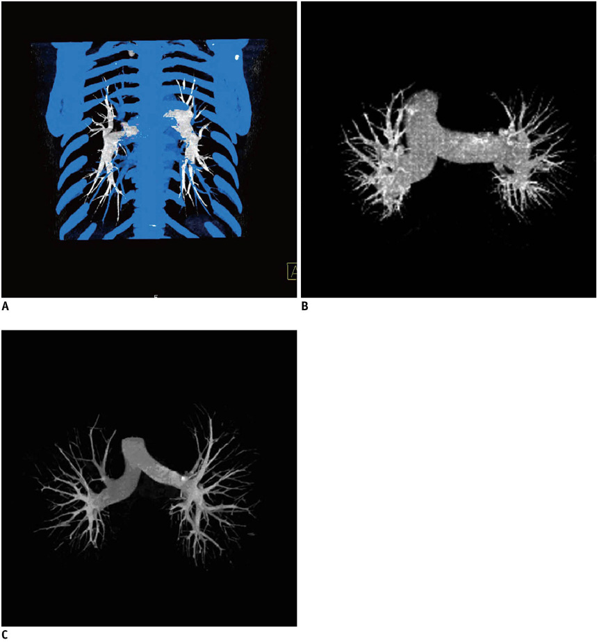
Fig. 1 Bone and pulmonary veins were marked in blue and removed with bone removal tool (A). 3D pulmonary artery trees from patients with pulmonary hypertension (B) and without pulmonary hypertension (C) were obtained after vessel segmentation.
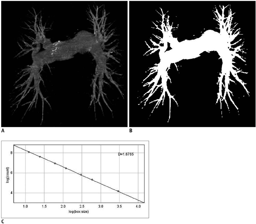
Fig. 2 Coronal projection of 2D pulmonary artery from patient with pulmonary hypertension (A), binary view of 2D projection (B) and graph of fractal dimension was obtained by box-counting method (C).
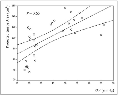
Fig. 3 Pulmonary arterial pressure (PAP) was plotted against projected image area of pulmonary arteries. There was correlation of pulmonary arterial pressure with projected image area (r = 0.65, p < 0.05, Pearson).
Reference
-
1. D'Alonzo GE, Barst RJ, Ayres SM, Bergofsky EH, Brundage BH, Detre KM, et al. Survival in patients with primary pulmonary hypertension. Results from a national prospective registry. Ann Intern Med. 1991. 115:343–349.2. Schannwell CM, Steiner S, Strauer BE. Diagnostics in pulmonary hypertension. J Physiol Pharmacol. 2007. 58:Suppl 5. 591–602.3. Arcasoy SM, Christie JD, Ferrari VA, Sutton MS, Zisman DA, Blumenthal NP, et al. Echocardiographic assessment of pulmonary hypertension in patients with advanced lung disease. Am J Respir Crit Care Med. 2003. 167:735–740.4. Sciomer S, Badagliacca R, Fedele F. Pulmonary hypertension: echocardiographic assessment. Ital Heart J. 2005. 6:840–845.5. Edwards PD, Bull RK, Coulden R. CT measurement of main pulmonary artery diameter. Br J Radiol. 1998. 71:1018–1020.6. Heinrich M, Uder M, Tscholl D, Grgic A, Kramann B, Schafers HJ. CT scan findings in chronic thromboembolic pulmonary hypertension: predictors of hemodynamic improvement after pulmonary thromboendarterectomy. Chest. 2005. 127:1606–1613.7. Kuriyama K, Gamsu G, Stern RG, Cann CE, Herfkens RJ, Brundage BH. CT-determined pulmonary artery diameters in predicting pulmonary hypertension. Invest Radiol. 1984. 19:16–22.8. Ng CS, Wells AU, Padley SP. A CT sign of chronic pulmonary arterial hypertension: the ratio of main pulmonary artery to aortic diameter. J Thorac Imaging. 1999. 14:270–278.9. Tan RT, Kuzo R, Goodman LR, Siegel R, Haasler GB, Presberg KW. Medical College of Wisconsin Lung Transplant Group. Utility of CT scan evaluation for predicting pulmonary hypertension in patients with parenchymal lung disease. Chest. 1998. 113:1250–1256.10. Maggi F, Winterwerp JC. Method for computing the three-dimensional capacity dimension from two-dimensional projections of fractal aggregates. Phys Rev E Stat Nonlin Soft Matter Phys. 2004. 69:011405.11. Weibel ER. Fractal geometry: a design principle for living organisms. Am J Physiol. 1991. 261:L361–L369.12. Goh V, Sanghera B, Wellsted DM, Sundin J, Halligan S. Assessment of the spatial pattern of colorectal tumour perfusion estimated at perfusion CT using two-dimensional fractal analysis. Eur Radiol. 2009. 19:1358–1365.13. Kamiya A, Takahashi T. Quantitative assessments of morphological and functional properties of biological trees based on their fractal nature. J Appl Physiol. 2007. 102:2315–2323.14. Bankier AA, Madani A, Gevenois PA. CT quantification of pulmonary emphysema: assessment of lung structure and function. Crit Rev Comput Tomogr. 2002. 43:399–417.15. Boser SR, Park H, Perry SF, Menache MG, Green FH. Fractal geometry of airway remodeling in human asthma. Am J Respir Crit Care Med. 2005. 172:817–823.16. Kido S, Sasaki S. Fractal analysis for the assessment of pulmonary blood flow on chest radiographs. J Thorac Imaging. 2003. 18:80–86.17. Madani A, Keyzer C, Gevenois PA. Quantitative computed tomography assessment of lung structure and function in pulmonary emphysema. Eur Respir J. 2001. 18:720–730.18. Huang W, Yen RT, McLaurine M, Bledsoe G. Morphometry of the human pulmonary vasculature. J Appl Physiol. 1996. 81:2123–2133.19. Sciomer S, Magri D, Badagliacca R. Non-invasive assessment of pulmonary hypertension: Doppler-echocardiography. Pulm Pharmacol Ther. 2007. 20:135–140.20. Kido S, Kuriyama K, Higashiyama M, Kasugai T, Kuroda C. Fractal analysis of internal and peripheral textures of small peripheral bronchogenic carcinomas in thin-section computed tomography: comparison of bronchioloalveolar cell carcinomas with nonbronchioloalveolar cell carcinomas. J Comput Assist Tomogr. 2003. 27:56–61.21. Wu YT, Shyu KK, Jao CW, Wang ZY, Soong BW, Wu HM, et al. Fractal dimension analysis for quantifying cerebellar morphological change of multiple system atrophy of the cerebellar type (MSA-C). Neuroimage. 2010. 49:539–551.22. Lv D, Guo X, Wang X, Zhang J, Fang J. Computerized characterization of prostate cancer by fractal analysis in MR images. J Magn Reson Imaging. 2009. 30:161–168.23. Kim N, Seo JB, Song KS, Chae EJ, Kang SH. Semi-automatic measurement of the airway dimension by computed tomography using the full-with-half-maximum method: a study of the measurement accuracy according to the orientation of an artificial airway. Korean J Radiol. 2008. 9:236–242.24. Kim N, Seo JB, Song KS, Chae EJ, Kang SH. Semi-automatic measurement of the airway dimension by computed tomography using the full-width-half-maximum method: a study on the measurement accuracy according to the CT parameters and size of the airway. Korean J Radiol. 2008. 9:226–235.25. Haimovici JB, Trotman-Dickenson B, Halpern EF, Dec GW, Ginns LC, Shepard JA, et al. Relationship between pulmonary artery diameter at computed tomography and pulmonary artery pressures at right-sided heart catheterization. Massachusetts General Hospital Lung Transplantation Program. Acad Radiol. 1997. 4:327–333.26. Froelich JJ, Koenig H, Knaak L, Krass S, Klose KJ. Relationship between pulmonary artery volumes at computed tomography and pulmonary artery pressures in patients with- and without pulmonary hypertension. Eur J Radiol. 2008. 67:466–471.27. Ley S, Mereles D, Risse F, Grunig E, Ley-Zaporozhan J, Tecer Z, et al. Quantitative 3D pulmonary MR-perfusion in patients with pulmonary arterial hypertension: correlation with invasive pressure measurements. Eur J Radiol. 2007. 61:251–255.28. Boxt LM, Katz J, Liebovitch LS, Jones R, Esser PD, Reid L. Fractal analysis of pulmonary arteries: the fractal dimension is lower in pulmonary hypertension. J Thorac Imaging. 1994. 9:8–13.29. Molthen RC, Karau KL, Dawson CA. Quantitative models of the rat pulmonary arterial tree morphometry applied to hypoxia-induced arterial remodeling. J Appl Physiol. 2004. 97:2372–2384. discussion 2354.30. Fernández E, Jelinek HF. Use of fractal theory in neuroscience: methods, advantages, and potential problems. Methods. 2001. 24:309–321.31. Kassab GS. Scaling laws of vascular trees: of form and function. Am J Physiol Heart Circ Physiol. 2006. 290:H894–H903.
- Full Text Links
-
- Actions
-
Cited
- CITED
-
- Close
- Share
-
- Similar articles
-
- Fractal Dimension Of Ct Images Of Normal Parotid Glands
- A Case of Giant Pulmonary Artery Aneurysm with Severe Pulmonary Hypertension
- Comparatives Study of Pulmonary Artery and Pulmonary Venous Wedge Pressure in Congenital Heart Disease
- Clinical Year in Review of Pulmonary Vascular Disease
- A Case of Pulmonary Hypertension Recurred by Graves' Disease
