Korean J Radiol.
2010 Dec;11(6):697-700. 10.3348/kjr.2010.11.6.697.
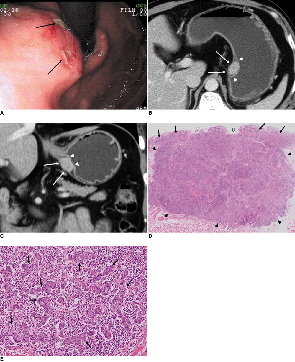
Epstein-Barr Virus-Associated Lymphoepithelioma-Like Gastric Carcinoma Presenting as a Submucosal Mass: CT Findings with Pathologic Correlation
- Affiliations
-
- 1Department of Radiology, Cheonan Hospital, Soonchunhyang University, Cheonan 330-721, Korea. rad2000@hanmail.net
- 2Department of Pathology, Cheonan Hospital, Soonchunhyang University, Cheonan 330-721, Korea.
- 3Department of Internal Medicine, Cheonan Hospital, Soonchunhyang University, Cheonan 330-721, Korea.
- KMID: 1119235
- DOI: http://doi.org/10.3348/kjr.2010.11.6.697
Abstract
- A lymphoepithelioma-like carcinoma, characterized by a carcinoma with heavy lymphocyte infiltration, is one of the histological patterns observed in patients with Epstein-Barr virus (EBV)-associated gastric carcinoma. Less than half of invasive carcinomas with lymphoepithelioma-like histology can grow to make a submucosal mass. These tumors generally have a better prognosis than conventional adenocarcinomas. We report a case of an EBV-associated lymphoepithelioma-like gastric carcinoma that presented as a submucosal mass on multi-detector (MD) CT and correlate them with the pathology.
MeSH Terms
Figure
Reference
-
1. Arikawa J, Tokunaga M, Satoh E, Tanaka S, Land CE. Morphological characteristics of Epstein-Barr virus-related early gastric carcinoma: a case-control study. Pathol Int. 1997. 47:360–367.2. Herath CH, Chetty R. Epstein-Barr virus-associated lymphoepithelioma-like gastric carcinoma. Arch Pathol Lab Med. 2008. 132:706–709.3. Murphy G, Pfeiffer R, Camargo MC, Rabkin CS. Meta-analysis shows that prevalence of Epstein-Barr virus-positive gastric cancer differs based on sex and anatomic location. Gastroenterology. 2009. 137:824–833.4. Lee JH, Kim SH, Han SH, An JS, Lee ES, Kim YS. Clinicopathological and molecular characteristics of Epstein-Barr virus-associated gastric carcinoma: a meta-analysis. J Gastroenterol Hepatol. 2009. 24:354–365.5. Burke AP, Yen TS, Shekitka KM, Sobin LH. Lymphoepithelial carcinoma of the stomach with Epstein-Barr virus demonstrated by polymerase chain reaction. Mod Pathol. 1990. 3:377–380.6. Iezzoni JC, Gaffey MJ, Weiss LM. The role of Epstein-Barr virus in lymphoepithelioma-like carcinomas. Am J Clin Pathol. 1995. 103:308–315.7. Nishikawa J, Yanai H, Mizugaki Y, Takada K, Tada M, Okita K. Case report: hypoechoic submucosal nodules: a sign of Epstein-Barr virus-associated early gastric cancer. J Gastroenterol Hepatol. 1998. 13:585–590.8. Shah KM, Young LS. Epstein-Barr virus and carcinogenesis: beyond Burkitt's lymphoma. Clin Microbiol Infect. 2009. 15:982–988.9. Song HJ, Srivastava A, Lee J, Kim YS, Kim KM, Ki Kang W, et al. Host inflammatory response predicts survival of patients with Epstein-Barr virus-associated gastric carcinoma. Gastroenterology. 2010. 139:84–92.10. Wu MS, Shun CT, Wu CC, Hsu TY, Lin MT, Chang MC, et al. Epstein-Barr virus-associated gastric carcinomas: relation to H. pylori infection and genetic alterations. Gastroenterology. 2000. 118:1031–1038.11. Maeda E, Akahane M, Uozaki H, Kato N, Hayashi N, Fukayama M, et al. CT appearance of Epstein-Barr virus-associated gastric carcinoma. Abdom Imaging. 2009. 34:618–625.12. Yanai H, Nishikawa J, Mizugaki Y, Shimizu N, Takada K, Matsusaki K, et al. Endoscopic and pathologic features of Epstein-Barr virus-associated gastric carcinoma. Gastrointest Endosc. 1997. 45:236–242.
- Full Text Links
- Actions
-
Cited
- CITED
-
- Close
- Share
- Similar articles
-
- A Case of Gastric Lymphoepithelioma-like Carcinoma Presenting as a Submucosal Tumor
- Pathology of Epstein-Barr Virus-Associated Gastric Carcinoma and Its Relationship to Prognosis
- A Case of Lymphoepithelioma-like Carcinoma Arising from the Parotid Gland
- A case of submucosal gastric lymphoepithelioma-like carcinoma
- Gastric Lymphoepithelioma-like Carcinoma Diagnosed and Treated by Endoscopic Submucosal Dissection: Review of the Literature


