Korean J Radiol.
2010 Dec;11(6):632-639. 10.3348/kjr.2010.11.6.632.
Value of Power Doppler and Gray-Scale US in the Diagnosis of Carpal Tunnel Syndrome: Contribution of Cross-Sectional Area just before the Tunnel Inlet as Compared with the Cross-Sectional Area at the Tunnel
- Affiliations
-
- 1Department of Radiology, Osmangazi University Hospital, Turkey. nevbahara@yahoo.com
- 2Department of Neurology, Osmangazi University Hospital, Turkey.
- 3Department of Physical Therapy and Rehabilitation, Osmangazi University Hospital, Turkey.
- KMID: 1119226
- DOI: http://doi.org/10.3348/kjr.2010.11.6.632
Abstract
OBJECTIVE
To determine the value of gray-scale and power Doppler ultrasonography in the evaluation of carpal tunnel syndrome (CTS).
MATERIALS AND METHODS
Median nerves at the carpal tunnel were evaluated by using gray-scale and power Doppler ultrasonography and by using accepted and new criteria in 42 patients with CTS (62 wrists) confirmed by electromyogram and 33 control subjects. We evaluated the cross-sectional area of the nerve just proximal to the tunnel inlet (CSAa), and at mid level (CSAb). We then calculated the percentage area increase of CSAb, and area difference (CSAb-CSAa). We measured two dimensions of the nerve at the distal level to calculate the flattening ratio. The power Doppler ultrasonography was used to assess the number of vessels, which proceeded to give a score according to the vessel number, and lastly evaluated the statistical significance by comparing the means of patients with control subjects by the Student t test for independent samples. Sensitivities and specificities were determined for sonographic characteristics mentioned above. We obtained the receiver operating characteristic (ROC) curve to assess the optimal cut-off values for the diagnosis of CTS.
RESULTS
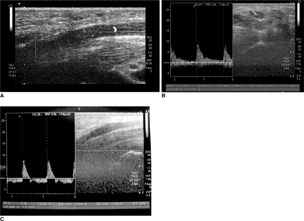
A statistically significant difference was found between patients and the control group for mean CSAb, area difference, percentage area increase, and flattening ratio (p < 0.001, p < 0.001, p < 0.001, p < 0.05, respectively). From the ROC curve we obtained optimal cut-off values of 11 mm2 for CSAb, 3.65 for area difference, 50% for the percentage of area increase, and 2.6 for the flattening ratio. The mean number of vessels obtained by power Doppler ultrasonography from the median nerve was 1.2. We could not detect vessels from healthy volunteers. Mean CSAbs related to vascularity intensity scores were as follows: score 0: 12.3 +/- 2.8 mm2, score 1: 12.3 +/- 3.1 mm2, score 2: 14.95 +/- 3.5 mm2, score 3: 19.3 +/- 3.8 mm2. The mean PI value in vessels of the median nerve was 4.1 +/- 1.
CONCLUSION
Gray-scale and power Doppler ultrasonography are useful in the evaluation of CTS.
MeSH Terms
Figure
Cited by 1 articles
-
Use of Magnetic Resonance Neurography for Evaluating the Distribution and Patterns of Chronic Inflammatory Demyelinating Polyneuropathy
Xiaoyun Su, Xiangquan Kong, Zuneng Lu, Min Zhou, Jing Wang, Xiaoming Liu, Xiangchuang Kong, Huiting Zhang, Chuansheng Zheng
Korean J Radiol. 2020;21(4):483-493. doi: 10.3348/kjr.2019.0739.
Reference
-
1. Duncan I, Sullivan P, Lomas F. Sonography in the diagnosis of carpal tunnel syndrome. AJR Am J Roentgenol. 1999. 173:681–684.2. Sarría L, Cabada T, Cozcolluela R, Martínez-Berganza T, García S. Carpal tunnel syndrome: usefulness of sonography. Eur Radiol. 2000. 10:1920–1925.3. Chen P, Maklad N, Redwine M, Zelitt D. Dynamic high-resolution sonography of the carpal tunnel. AJR Am J Roentgenol. 1997. 168:533–537.4. Klauser AS, Halpern EJ, De Zordo T, Feuchtner GM, Arora R, Gruber J, et al. Carpal tunnel syndrome assessment with US: value of additional cross-sectional area measurements of the median nerve in patients versus healthy volunteers. Radiology. 2009. 250:171–177.5. Buchberger W, Judmaier W, Birbamer G, Lener M, Schmidauer C. Carpal tunnel syndrome: diagnosis with high-resolution sonography. AJR Am J Roentgenol. 1992. 159:793–798.6. Ziswiler HR, Reichenbach S, Vögelin E, Bachmann LM, Villiger PM, Jüni P. Diagnostic value of sonography in patients with suspected carpal tunnel syndrome: a prospective study. Arthritis Rheum. 2005. 52:304–311.7. Wong SM, Griffith JF, Hui AC, Lo SK, Fu M, Wong KS. Carpal tunnel syndrome: diagnostic usefulness of sonography. Radiology. 2004. 232:93–99.8. Keberle M, Jenett M, Kenn W, Reiners K, Peter M, Haerten R, et al. Technical advances in ultrasound and MR imaging of carpal tunnel syndrome. Eur Radiol. 2000. 10:1043–1050.9. El Miedany YM, Aty SA, Ashour S. Ultrasonography versus nerve conduction study in patients with carpal tunnel syndrome: substantive or complementary tests? Rheumatology (Oxford). 2004. 43:887–895.10. Visser LH, Smidt MH, Lee ML. High-resolution sonography versus EMG in the diagnosis of carpal tunnel syndrome. J Neurol Neurosurg Psychiatry. 2008. 79:63–67.11. Keleş I, Karagülle Kendi AT, Aydin G, Zöğ SG, Orkun S. Diagnostic precision of ultrasonography in patients with carpal tunnel syndrome. Am J Phys Med Rehabil. 2005. 84:443–450.12. Leonard L, Rangan A, Doyle G, Taylor G. Carpal tunnel syndrome - is high-frequency ultrasound a useful diagnostic tool? J Hand Surg Br. 2003. 28:77–79.13. Ashraf AR, Jali R, Moghtaderi AR, Yazdani AH. The diagnostic value of ultrasonography in patients with electrophysiologicaly confirmed carpal tunnel syndrome. Electromyogr Clin Neurophysiol. 2009. 49:3–8.14. Mallouhi A, Pültzl P, Trieb T, Piza H, Bodner G. Predictors of carpal tunnel syndrome: accuracy of gray-scale and color Doppler sonography. AJR Am J Roentgenol. 2006. 186:1240–1245.15. Radack DM, Schweitzer ME, Taras J. Carpal tunnel syndrome: are the MR findings a result of population selection bias? AJR Am J Roentgenol. 1997. 169:1649–1653.16. Klauser A, Frauscher F, Schirmer M, Halpern E, Pallwein L, Herold M, et al. The value of contrast-enhanced color Doppler ultrasound in the detection of vascularization of finger joints in patients with rheumatoid arthritis. Arthritis Rheum. 2002. 46:647–653.17. Shio K, Homma F, Kanno Y, Yamadera Y, Ohguchi Y, Nishimaki T, et al. Doppler sonographic comparative study on usefulness of synovial vascularity between knee and metacarpophalangeal joints for evaluation of articular inflammation in patients with rheumatoid arthritis treated by infliximab. Mod Rheumatol. 2006. 16:220–225.18. Cho KH, Lee SM, Lee YH, Suh KJ. Ultrasound diagnosis of either an occult or missed fracture of an extremity in pediatric-aged children. Korean J Radiol. 2010. 11:84–94.19. Lee D, van Holsbeeck MT, Janevski PK, Ganos DL, Ditmars DM, Darian VB. Diagnosis of carpal tunnel syndrome. Ultrasound versus electromyography. Radiol Clin North Am. 1999. 37:859–872.20. Sucher BM. Ultrasound imaging of the carpal tunnel during median nerve compression. Curr Rev Musculoskelet Med. 2009. 2:134–146.21. Cartwright MS, Shin HW, Passmore LV, Walker FO. Ultrasonographic reference values for assessing the normal median nerve in adults. J Neuroimaging. 2009. 19:47–51.22. Elwakil TF, Elazzazi A, Shokeir H. Treatment of carpal tunnel syndrome by low-level laser versus open carpal tunnel release. Lasers Med Sci. 2007. 22:265–270.23. Kook SH, Park HW, Lee YR, Lee YU, Pae WK, Park YL. Evaluation of solid breast lesions with power Doppler sonography. J Clin Ultrasound. 1999. 27:231–237.24. Birdwell RL, Ikeda DM, Jeffrey SS, Jeffrey RB Jr. Preliminary experience with power Doppler imaging of solid breast masses. AJR Am J Roentgenol. 1997. 169:703–707.25. Lee SH, Suh JS, Shin MJ, Kim SM, Kim N, Suh SH. Quantitative assessment of synovial vascularity using contrast-enhanced power Doppler ultrasonography: correlation with histologic findings and MR imaging findings in arthritic rabbit knee model. Korean J Radiol. 2008. 9:45–53.
- Full Text Links
- Actions
-
Cited
- CITED
-
- Close
- Share
- Similar articles
-
- RE: Value of Power Doppler and Gray-Scale US in the Diagnosis of Carpal Tunnel Syndrome: Contribution of Cross-Sectional Area just before the Tunnel Inlet as Compared with the Cross-Sectional Area at the Tunnel
- The Abnormal Ultrasonographic Findings of Carpal Tunnel in Carpal Tunnel Syndrome
- Analysis of Sonographic Measurement by Anatomical Area in Carpal Tunnel Syndrome and Correlation the Measurement with Electrodiagnostic Study
- Ultrasonographic Study of Median Nerve According to Changed Wrist Position in Diabetics and Normal Subjects
- Ultrasonographic Findings of Mild and Very Mild Carpal Tunnel Syndrome


