Korean J Ophthalmol.
2004 Jun;18(1):1-8. 10.3341/kjo.2004.18.1.1.
Ability of Scanning Laser Polarimetry (GDx) to Discriminate among Early Glaucomatous, Ocular Hypertensive and Normal Eyes in the Korean Population
- Affiliations
-
- 1Department of Ophthalmology, University of Ulsan, College of Medicine, Asan Medical Center, Korea.
- 2Dr. Ha's Eye Clinic, Seoul, Korea.
- KMID: 1115776
- DOI: http://doi.org/10.3341/kjo.2004.18.1.1
Abstract
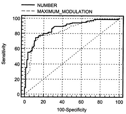
- We investigated the ability of the GDx-Nerve Fiber Analyzer (NFA) to discriminate between normal and early glaucomatous eyes among Korean individuals by reviewing the medical records of 217 consecutive subjects: 61 early glaucoma patients, 68 ocular hypertensive patients, and 88 normal subjects. GDx parameters were compared using ANOVA. The Receiver Operating Characteristics (ROC) curve for each GDx-NFA variable was used to diagnose each parameter, and Pearson correlation coefficients were calculated to assess the association between GDx-NFA parameters and visual field indices in early glaucoma. The best GDx parameters to discriminate between early glaucomatous and normal subjects were the number, maximum modulation, ellipse modulation and inferior ratio (i.e. area under the ROC curve > 0.8). A value for the Number of equal to or greater than 27 was optimal for detecting early glaucoma, with a sensitivity of 80.3% and specificity of 80.7%. In addition, symmetry was positively correlated with the corrected pattern standard deviation (CPSD) among visual field indices in early glaucoma.
MeSH Terms
-
*Diagnostic Techniques, Ophthalmological
Female
Glaucoma, Open-Angle/*diagnosis/ethnology
Humans
Intraocular Pressure
Korea/epidemiology
Male
Middle Aged
Nerve Fibers/*pathology
Ocular Hypertension/diagnosis/ethnology
Optic Nerve Diseases/*diagnosis/ethnology
ROC Curve
Retinal Ganglion Cells/*pathology
Retrospective Studies
Sensitivity and Specificity
Visual Fields
Figure
Cited by 1 articles
-
Longitudinal Analysis of Retinal Nerve Fiber Layer Thickness With GDx-VCC in Glaucoma Suspect
Wool Suh, Roo-Min Jun, Kyu-Ryong Choi
J Korean Ophthalmol Soc. 2009;50(2):235-241. doi: 10.3341/jkos.2009.50.2.235.
Reference
-
1. Dreher AW, Reiter K, Weinreb SN. Spatially resolved birefringence of the retinal nerve fiber layer assessed with a retinal laser ellipsometer. Appl Opt. 1992. 31:3730–3735.2. Dreher AW, Reiter K. Retinal laser ellipsometry: a new method for measuring the retinal nerve fiber layer thickness distribution. Clin Vision Sci. 1992. 7:481–488.3. Weinreb RN, Lusky M, Bartsch DU, Morsman D. Effect of repetitive imaging on topographic measurements of the optic nerve head. Arch Ophthalmol. 1993. 111:636–638.4. Weinreb RN, Dreher AW, Coleman A, Quigley H, Shaw B, Reiter K. Histopathologic validation of Fourier-ellipsometry measurements of retinal nerve fiber layer thickness. Arch Ophthalmol. 1990. 108:557–560.5. Greenfield DS, Knighton RW, Huang XR. Effect of corneal polarization axis on assessment of retinal nerve fiber layer thickness by scanning laser polarimetry. Am J Ophthalmol. 2000. 129:715–722.6. Chi QM, Tomita G, Inazumi K, Hayakawa T, Ido T, Kitazawa Y. Evaluation of the effect of ageing on the retinal nerve fiber layer thickness using scanning laser polarimetry. J Glaucoma. 1995. 4:406–413.7. Bone RA. The role of the macular pigment in the detection of polarized light. Vision Res. 1980. 20:213–220.8. Hoh ST, Greenfield DS, Liebmann JM, Maw R, Ishikawa H, Chew SJ, Ritch R. Factors affecting image acquisition during scanning laser polarimetry. Ophthalmic Surg Lasers. 1998. 29:545–551.9. Tjon-Fo-Sang MJ, Lemij HG. The sensitivity and specificity of nerve fiber layer measurements in glaucoma as determined with scanning laser polarimetry. Am J Ophthalmol. 1997. 123:62–69.10. Zangwill LM, Bowd C, Berry CC, Williams J, Blumenthal EZ, Sanchez-Galeana CA, Vasile C, Weinreb RN. Discriminating between normal and glaucomatous eyes using the Heidelberg Retina Tomograph, GDx Nerve Fiber Analyzer, and Optical Coherence Tomograph. Arch Ophthalmol. 2001. 119:985–993.11. Weinreb RN, Zangwill LM, Berry CC, Bathija R, Sample PA. Detection of glaucoma with scanning laser polarimetry. Arch Ophthalmol. 1998. 116:1583–1589.12. Funaki S, Shirakashi M, Yaoeda K, Abe H, Kunimatsu H, Suzuki Y, Tomita G, Araie M, Yamada N, Uchida H, Yamamoto T, Kitazawa Y. Specificity and sensitivity of glaucoma detection in the Japanese population using scanning laser polarimetry. Br J Ophthalmol. 2002. 86:70–74.13. Kook MS, Sung K, Park RH, Kim KR, Kim ST, Kang W. Reproducibility of scanning laser polarimetry (GDx) of peripapillary retinal nerve fiber layer thickness in normal subjects. Graefe's Arch Clin Exp Ophthalmol. 2001. 239:118–121.14. Caprioli J. Discrimination between normal and glaucomatous eyes. Invest Ophthalmol Vis Sci. 1992. 33:153–159.15. Katz J, Tielsch JM, Quigley HA, Javitt J, Witt K, Sommer A. Automated suprathreshold screening for glaucoma: the Baltimore Eye Survey. Invest Ophthalmol Vis Sci. 1993. 34:3271–3277.16. Kosoko O, Sommer A, Auer C. Screening with automated perimetry using a threshold-related three-level algorithm. Ophthalmology. 1986. 93:882–886.17. DeLong ER, DeLong DM, Clarke-Pearson DL. Comparing the areas under two or more correlated receiver operating characteristic curves: a nonparametric approach. Biometrics. 1988. 44:837–845.18. Quigley HA, Addicks EM, Green WR. Optic nerve damage in human glaucoma. III. Quantitative correlation of nerve fiber loss and visual field defect in glaucoma, ischemic neuropathy, papilledema, and toxic neuropathy. Arch Ophthalmol. 1982. 100:135–146.19. Provis JM, van Driel D, Billson FA, Russell P. Human fetal optic nerve: overproduction and elimination of retinal axons during development. J Comp Neurol. 1985. 238:92–100.20. Quigley HA, Brown AE, Morrison JD, Drance SM. The size and shape of the optic disc in normal human eyes. Arch Ophthalmol. 1990. 108:51–57.21. Weinreb RN, Shakiba S, Sample PA, Shahrokni S, van Horn S, Garden VS, Asawaphureekorn S, Zangwill L. Association between quantitative nerve fiber layer measurement and visual field loss in glaucoma. Am J Ophthalmol. 1995. 120:732–738.22. Choplin NT, Lundy DC, Dreher AW. Differentiating patients with glaucoma from glaucoma suspects and normal subjects by nerve fiber layer assessment with scanning laser polarimetry. Ophthalmology. 1998. 105:2068–2076.23. Kwon YH, Hong S, Honkanen RA, Alward WLM. Correlation of automated visual field parameters and peripapillary nerve fiber layer thickness as measured by scanning laser polarimetry. J Glaucoma. 2000. 9:281–288.24. Paczka JA, Friedman DS, Quigley HA, Barron Y, Vitale S. Diagnostic capabilities of frequency-doubling technology, scanning laser polarimetry, and nerve fiber layer photographs to distinguish glaucomatous damage. Am J Ophthalmol. 2001. 131:188–197.
- Full Text Links
- Actions
-
Cited
- CITED
-
- Close
- Share
- Similar articles
-
- GDx-VCC Performance to Discriminate Normal, Pre-perimetric Glaucomatous Eyes
- Diagnostic Ability of Scanning Laser Polarimetry with Enhanced Corneal Compensation in the Eye with Typical and Atypical Retadation Pattern
- Scanning Laser Polarimetry Using Variable Corneal Compensation in Detection of Localized Visual Field Defects
- Discriminating Ability of Scanning Laser Polarimetry with Variable Corneal Compensation in Normal and Glaucomatous Eyes
- Influence of Lens Opacity on Nerve Fiber Layer Analysis in Glaucomatous and Normal Eyes


