Korean J Ophthalmol.
2011 Apr;25(2):77-83. 10.3341/kjo.2011.25.2.77.
Changes in Anterior Chamber Configuration after Cataract Surgery as Measured by Anterior Segment Optical Coherence Tomography
- Affiliations
-
- 1Department of Ophthalmology, Seoul National University Bundang Hospital, Seongnam, Korea.
- 2Department of Ophthalmology, Seoul National University Hospital, Seoul National University College of Medicine, Seoul, Korea. kihopark@snu.ac.kr
- KMID: 1111867
- DOI: http://doi.org/10.3341/kjo.2011.25.2.77
Abstract
- PURPOSE
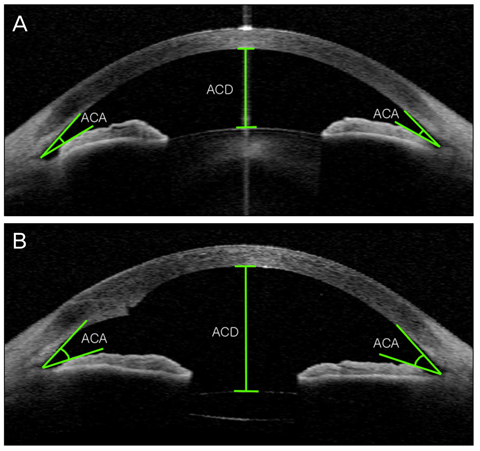
To evaluate the changes in anterior chamber depth (ACD) and angle width induced by phacoemulsification and intraocular lens (IOL) implantation in normal eyes using anterior segment optical coherence tomography (AS-OCT).
METHODS
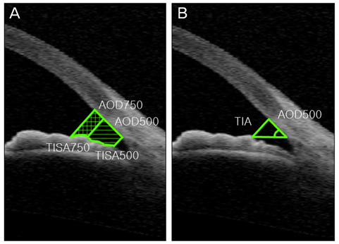
Forty-five eyes (45 patients) underwent AS-OCT imaging to evaluate anterior chamber configuration before and 2 days after phacoemulsification and IOL implantation. We analyzed the central ACD and angle width using different methods: anterior chamber angle (ACA), trabecular-iris angle (TIA), angle opening distance (AOD), and trabecular iris surface area (TISA) in the nasal and temporal quadrants. Comparison between preoperative and postoperative measurement was done using paired t-tests and each of the angle parameters was analyzed with Pearson correlation testing. Subgroup analyses according to the IOL and axial length were performed with a general multivariate linear model adjusted for age.
RESULTS
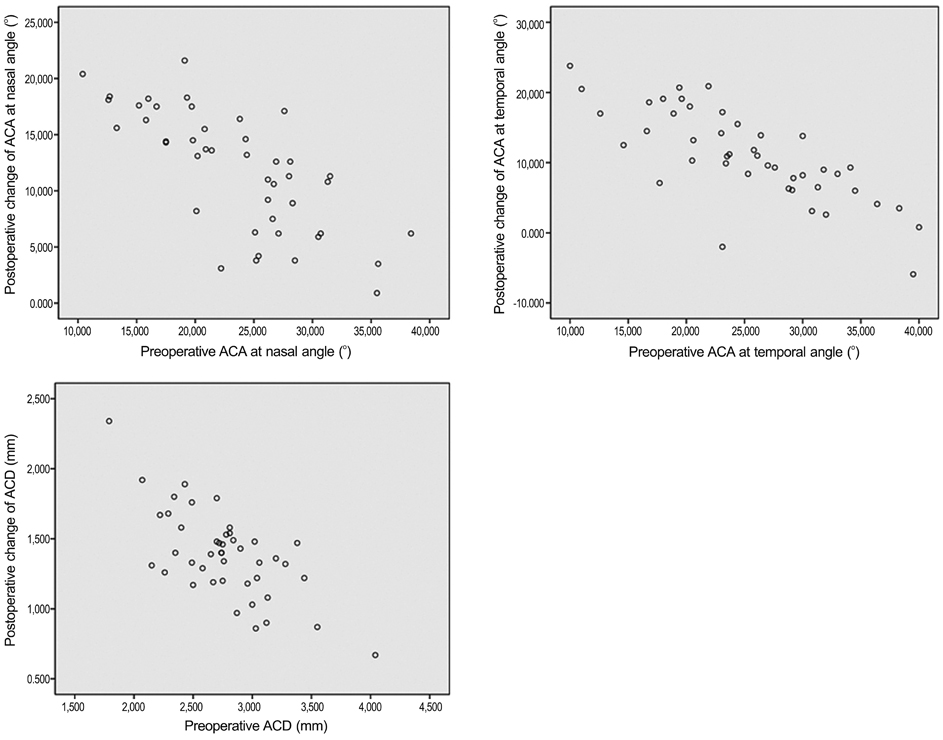
Before surgery, the mean anterior chamber angle widths were 23.21 +/- 6.70degrees in the nasal quadrant and 24.89 +/- 7.66degrees in the temporal quadrant. The mean central ACD was 2.75 +/- 0.43 mm. After phacoemulsification and IOL implantation, the anterior chamber angle width increased significantly to 35.16 +/- 4.65degrees in the nasal quadrant (p = 0.001) and 36.03 +/- 4.86degrees in the temporal quadrant (p = 0.001). Also, central ACD increased to 4.14 +/- 0.31 mm (p = 0.001). AOD, TISA, and TIA increased significantly after cataract surgery and showed positive correlation with ACA.
CONCLUSIONS
After cataract surgery, the ACD and angle width significantly increased in eyes with cataract. AS-OCT is a good method for obtaining quantitative data regarding anterior chamber configuration.
MeSH Terms
Figure
Cited by 1 articles
-
Anterior Chamber and Lens Position before and after Phacoemulsification According to Axial Length
Suk Hoon Jung, Seonjoo Kim, So Hyang Chung
J Korean Ophthalmol Soc. 2020;61(1):17-26. doi: 10.3341/jkos.2020.61.1.17.
Reference
-
1. Boker T, Sheqem J, Rauwolf M, Wegener A. Anterior chamber angle biometry: a comparison of Scheimpflug photography and ultrasound biomicroscopy. Ophthalmic Res. 1995. 27:Suppl 1. 104–109.2. Dada T, Sihota R, Gadia R, et al. Comparison of anterior segment optical coherence tomography and ultrasound biomicroscopy for assessment of the anterior segment. J Cataract Refract Surg. 2007. 33:837–840.3. Kucumen RB, Yenerel NM, Gorgun E, et al. Anterior segment optical coherence tomography measurement of anterior chamber depth and angle changes after phacoemulsification and intraocular lens implantation. J Cataract Refract Surg. 2008. 34:1694–1698.4. Lam AK, Chan R, Woo GC, et al. Intra-observer and inter-observer repeatability of anterior eye segment analysis system (EAS-1000) in anterior chamber configuration. Ophthalmic Physiol Opt. 2002. 22:552–559.5. Memarzadeh F, Tang M, Li Y, et al. Optical coherence tomography assessment of angle anatomy changes after cataract surgery. Am J Ophthalmol. 2007. 144:464–465.6. Pavlin CJ, Foster FS. Plateau iris syndrome: changes in angle opening associated with dark, light, and pilocarpine administration. Am J Ophthalmol. 1999. 128:288–291.7. Pavlin CJ, Ritch R, Foster FS. Ultrasound biomicroscopy in plateau iris syndrome. Am J Ophthalmol. 1992. 113:390–395.8. Pavlin CJ, Sherar MD, Foster FS. Subsurface ultrasound microscopic imaging of the intact eye. Ophthalmology. 1990. 97:244–250.9. Richards DW, Russell SR, Anderson DR. A method for improved biometry of the anterior chamber with a Scheimpflug technique. Invest Ophthalmol Vis Sci. 1988. 29:1826–1835.10. Sakuma T, Sawada A, Yamamoto T, Kitazawa Y. Appositional angle closure in eyes with narrow angles: an ultrasound biomicroscopic study. J Glaucoma. 1997. 6:165–169.11. Shibata T, Sasaki K, Sakamoto Y, Takahashi N. Quantitative chamber angle measurement utilizing image-processing techniques. Ophthalmic Res. 1990. 22:Suppl 1. 81–84.12. Nolan W. Anterior segment imaging: ultrasound biomicroscopy and anterior segment optical coherence tomography. Curr Opin Ophthalmol. 2008. 19:115–121.13. Radhakrishnan S, Goldsmith J, Huang D, et al. Comparison of optical coherence tomography and ultrasound biomicroscopy for detection of narrow anterior chamber angles. Arch Ophthalmol. 2005. 123:1053–1059.14. Radhakrishnan S, Huang D, Smith SD. Optical coherence tomography imaging of the anterior chamber angle. Ophthalmol Clin North Am. 2005. 18:375–381.15. Nolan W. Anterior segment imaging: identifying the landmarks. Br J Ophthalmol. 2008. 92:1575–1576.16. Radhakrishnan S, Rollins AM, Roth JE, et al. Real-time optical coherence tomography of the anterior segment at 1310 nm. Arch Ophthalmol. 2001. 119:1179–1185.17. Lee R, Ahmed II. Anterior segment optical coherence tomography: non-contact high resolution imaging of the anterior chamber. Tech Ophthalmol. 2006. 4:120–127.18. Li H, Leung CK, Cheung CY, et al. Repeatability and reproducibility of anterior chamber angle measurement with anterior segment optical coherence tomography. Br J Ophthalmol. 2007. 91:1490–1492.19. Mohamed S, Lee GK, Rao SK, et al. Repeatability and reproducibility of pachymetric mapping with Visante anterior segment-optical coherence tomography. Invest Ophthalmol Vis Sci. 2007. 48:5499–5504.20. Muller M, Dahmen G, Porksen E, et al. Anterior chamber angle measurement with optical coherence tomography: intraobserver and interobserver variability. J Cataract Refract Surg. 2006. 32:1803–1808.21. Radhakrishnan S, See J, Smith SD, et al. Reproducibility of anterior chamber angle measurements obtained with anterior segment optical coherence tomography. Invest Ophthalmol Vis Sci. 2007. 48:3683–3688.22. Hayashi K, Hayashi H, Nakao F, Hayashi F. Changes in anterior chamber angle width and depth after intraocular lens implantation in eyes with glaucoma. Ophthalmology. 2000. 107:698–703.23. Nolan WP, See JL, Aung T, et al. Changes in angle configuration after phacoemulsification measured by anterior segment optical coherence tomography. J Glaucoma. 2008. 17:455–459.24. Nonaka A, Kondo T, Kikuchi M, et al. Angle widening and alteration of ciliary process configuration after cataract surgery for primary angle closure. Ophthalmology. 2006. 113:437–441.25. Pereira FA, Cronemberger S. Ultrasound biomicroscopic study of anterior segment changes after phacoemulsification and foldable intraocular lens implantation. Ophthalmology. 2003. 110:1799–1806.26. Dawczynski J, Koenigsdoerffer E, Augsten R, Strobel J. Anterior segment optical coherence tomography for evaluation of changes in anterior chamber angle and depth after intraocular lens implantation in eyes with glaucoma. Eur J Ophthalmol. 2007. 17:363–367.27. Chang DH, Lee SC, Jin KH. Changes of anterior chamber depth and angle after cataract surgery measured by anterior segment OCT. J Korean Ophthalmol Soc. 2008. 49:1443–1452.28. Leung CK, Chan WM, Ko CY, et al. Visualization of anterior chamber angle dynamics using optical coherence tomography. Ophthalmology. 2005. 112:980–984.29. Leung CK, Cheung CY, Li H, et al. Dynamic analysis of dark-light changes of the anterior chamber angle with anterior segment OCT. Invest Ophthalmol Vis Sci. 2007. 48:4116–4122.30. Nemeth G, Vajas A, Tsorbatzoglou A, et al. Assessment and reproducibility of anterior chamber depth measurement with anterior segment optical coherence tomography compared with immersion ultrasonography. J Cataract Refract Surg. 2007. 33:443–447.31. Yi JH, Lee H, Hong S, et al. Anterior chamber measurements by pentacam and AS-OCT in eyes with normal open angles. Korean J Ophthalmol. 2008. 22:242–245.32. Dubbelman M, van der Heijde GL, Weeber HA. The thickness of the aging human lens obtained from corrected Scheimpflug images. Optom Vis Sci. 2001. 78:411–416.33. Williams DL. Lens morphometry determined by B-mode ultrasonography of the normal and cataractous canine lens. Vet Ophthalmol. 2004. 7:91–95.34. Kim JS, Shyn KH. Periodic biometry in three types of posteriorly implanted IOLs: PMMA, silicone, and acrylic soft, by EAS-1000 Scheimpflug photography. J Korean Ophthalmol Soc. 2000. 41:2205–2210.35. Hosny M, Alio JL, Claramonte P, et al. Relationship between anterior chamber depth, refractive state, corneal diameter, and axial length. J Refract Surg. 2000. 16:336–340.36. Pavlin CJ, Harasiewicz K, Foster FS. Ultrasound biomicroscopy of anterior segment structures in normal and glaucomatous eyes. Am J Ophthalmol. 1992. 113:381–389.
- Full Text Links
- Actions
-
Cited
- CITED
-
- Close
- Share
- Similar articles
-
- Changes of Anterior Chamber Depth and Angle After Cataract Surgery Measured by Anterior Segment OCT
- Changes in Anterior Chamber Depth and Angle After Phacoemulsification measured by Anterior Segment Optical Coherence Tomography
- Detection of Occludable Angles with the Pentacam and the Anterior Segment Optical Coherence Tomography
- Comparison of Anterior Segment Parameters Obtained by Anterior Segment Optical Coherence Tomography and Dual Rotating Scheimpflug Camera
- Comparison of Anterior Segment Measurements between Scheimpflug Camera and New Module of Optical Coherence Tomography


