Computed tomographic characteristics of acute thoracolumbar intervertebral disc disease in dogs
- Affiliations
-
- 1Department of Veterinary Medical Imaging, College of Veterinary Medicine, Seoul National University, Seoul 151-742, Korea. heeyoon@snu.ac.kr
- 2Department of Veterinary Surgery, College of Veterinary Medicine, Seoul National University, Seoul 151-742, Korea.
- KMID: 1106160
- DOI: http://doi.org/10.4142/jvs.2010.11.1.73
Abstract
- Forty canine patients with a presumptive diagnosis of the intervertebral disc herniation at the thoracolumbar region were imaged. A neurological examination was performed and all patients were classified under four grades by the examination. The degrees of attenuation of the herniated disc material were measured in Housefield units (HU) in each image. The ratio of the area to herniated disc material and the height to disc material were measured. The clinical grade was correlated with the area ratio of the herniated disc material to the spinal cord, but not correlated with the height ratio of that. In the patients with epidural hemorrhage at surgery, HUs of the herniated disc material was lower than those with no epidural hemorrhage at surgery. Non-contrast computed tomography scans of the spine can be useful in diagnosing acute intervertebral disc disease in chondrodystrophoid breeds, evaluating patient status and identifying concurrent epidural hemorrhage.
Keyword
MeSH Terms
Figure
-

Fig. 1 Location of disc herniation. The central (A), dorsal (B), left lateral (C), and right lateral (D) locations of disc herniation.

Fig. 2 Measurements for the index calculation with vertebra window (A) and spine window (B). 'a' represents the area of spinal canal and 'b' the area of herniated disc material, line 'cd' the height of spinal canal and line 'e-f' the height of herniated disc material. The formula for A-index calculation is b/a ×100 (%), and for H-index calculation (e to f / c to d) ×100 (%).
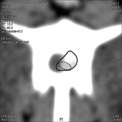
Fig. 3 Measurement of the HU of herniated disc materials. The solid line shows the total herniated disc area and the dotted line the region of interest of non-mineralized disc area.

Fig. 4 Mean A-index in relation to the grade by three different method of sampling (top, average of top 3 and average of top 5).
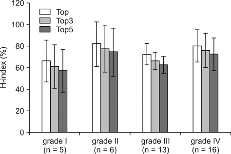
Fig. 5 Mean H-index in relation to the grade by three different methods of sampling (top, average of top 3 and average of top 5).
Cited by 1 articles
-
Computed tomographic evaluation of cervical vertebral canal and spinal cord morphometry in normal dogs
Eunjeong Seo, Jihye Choi, Mincheol Choi, Junghee Yoon
J Vet Sci. 2014;15(2):187-193. doi: 10.4142/jvs.2014.15.2.187.
Reference
-
1. Braund KG. Bojrab MJ, Smeak DD, Bloomberg MS, editors. Intervertebral disk disease. Disease Mechanisms in Small Animal Surgery. 1993. 2nd ed. Philadelphia: Lea & Febiger;960–970. .2. Bray JP, Burbidge HM. The canine intervertebral disk: part one: structure and function. J Am Anim Hosp Assoc. 1998. 34:55–63.
Article3. Coates JR. Intervertebral disk disease. Vet Clin North Am Small Anim Pract. 2000. 30:77–110.
Article4. Fagerlund MKJ, Thelander U, Friberg S. Size of lumbar disc hernias measured using computed tomography and related to sciatic symptoms. Acta Radiol. 1990. 31:555–558.
Article5. Goggin JE, Li AS, Franti CE. Canine intervertebral disk disease: Characterization by age, sex, breed, and anatomic site of involvement. Am J Vet Res. 1970. 31:1687–1692.6. Hoerlein BF. Intervertebral disc protrusions in the dog. I. Incidence and pathological lesions. Am J Vet Res. 1953. 14:260–283.7. Hoerlein BF. Canine Neurology: Diagnosis and Treatment. 1971. 2nd ed. Philadelphia: Saunders;307–391.8. Kirberger RM, Roos CJ, Lubbe AM. The radiological diagnosis of thoracolumbar disc disease in the Dachshund. Vet Radiol Ultrasound. 1992. 33:255–261.
Article9. Lamb CR. Common difficulties with myelographic diagnosis of acute intervertebral disc prolapse in the dog. J Small Anim Pract. 1994. 35:549–558.
Article10. Liptak JM, Watt PR, Thomson MJ, Copeland SE, Galloway AM. Hansen type I disk disease at T1-2 in a dachshund. Aust Vet J. 1999. 77:156–159.
Article11. Olby NJ, Muñana KR, Sharp NJH, Thrall DE. The computed tomographic appearance of acute thoracolumbar intervertebral disc herniations in dogs. Vet Radiol Ultrasound. 2000. 41:396–402.
Article12. Olsson SE. The dynamic factor in spinal cord compression: a study on dogs with special reference to cervical disc protrusions. J Neurosurg. 1958. 15:308–321.13. Resjö IM, Harwood-Nash DC, Fitz CR, Chuang S. Normal cord in infants and children examined with computed tomographic metrizamide myelography. Radiology. 1979. 130:691–696.
Article14. Sachsenheimer W, Hamer J, Müller HA. The value of spinal computed tomography in diagnosis of herniated lumbar discs. Acta Neurochir (Wien). 1982. 60:107–114.
Article15. Sharp NJH, Wheeler SJ. Small Animal Spinal Disorders: Diagnosis and Surgery. 2005. 2nd ed. Edinburgh: Elsevier;1–17. 121–135.16. Tarlov IM, Klinger H. Spinal cord compression studies. II. Time limits for recovery after acute compression in dogs. AMA Arch Neurol Psychiatry. 1954. 71:271–290.17. Thelander U, Fagerlund M, Friberg S, Larsson S. Describing the size of lumbar disc herniations using computed tomography: A comparison of different size index calculations and their relation to sciatica. Spine. 1994. 19:1979–1984.
Article18. Widmer WR, Thrall DE. Thrall DE, editor. Canine and feline intervertebral disc disease, myelography, and spinal cord disease. Textbook of Veterinary Diagnostic Radiology. 2002. 4th ed. Philadelphia: Saunders;110–133.
- Full Text Links
-
- Actions
-
Cited
- CITED
-
- Close
- Share
-
- Similar articles
-
- Mini-partial lateral corpectomy and hemilaminectomy for the treatment of heavily protruded thoracolumbar intervertebral disc in small dogs
- Transplantation of adipose derived mesenchymal stem cells for acute thoracolumbar disc disease with no deep pain perception in dogs
- Far lateral lumbar disc extrusion in a dachshund dog
- Preliminary study of presumptive intradural-intramedullary intervertebral disc extrusion in 20 dogs
- Biochemical Factors of Intervertebral Disc Degeneration: Implications for Disc Regeneration
