Stereolithographic biomodeling of equine ovary based on 3D serial digitizing device
- Affiliations
-
- 1College of Veterinary Medicine, Seoul National University, Seoul 151-742, Korea. kimura@snu.ac.kr
- 2RIKEN, Wako, Saitama, 351-0198, Japan.
- 3Obihiro University of Agriculture and Veterinary Medicine, Obihiro, Hokkaido, 08-8555, Japan.
- 4The Japan Racing Association, Tokyo, 320-0856, Japan.
- KMID: 1102974
- DOI: http://doi.org/10.4142/jvs.2009.10.2.161
Abstract
- The 3D internal structure microscopy (3D-ISM) was applied to the equine ovary, which possesses peculiar structural characteristics. Stereolithography was applied to make a life-sized model by means of data obtained from 3D-ISM. Images from serially sliced surfaces contributed to a successful 3D reconstruction of the equine ovary. Photopolymerized resin models of equine ovaries produced by stereolithography can clearly show the internal structure and spatial localizations in the ovary. The understanding of the spatial relationship between the ovulation fossa and follicles and/or corpora lutea in the equine ovary was a great benefit. The peculiar structure of the equine ovary could be thoroughly observed and understood through this model.
Keyword
MeSH Terms
Figure
-

Fig. 1 The computer simulation of the spatial arrangement of follicles in the equine ovary. These images were created by 3-dimensional reconstruction from serially sliced images. Assuming that the ovary is in the hexahedron box, six views are illustrated from the bottom surface (A), the top surface (B), the front surface (C), the back surface (D), the left side surface (E) and the right side surface (F). Follicles whose size is bigger than 10,000 mm3 marked in red, 3,000~9,999 mm3 marked in green, 1,000~2,999 mm3 marked in blue and 100~999 mm3 marked in white.
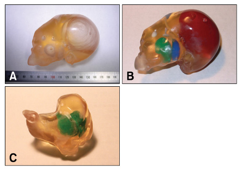
Fig. 2 The stereolithography product with empty spaces for the follicles (A), the follicles where the dye materials were injected (B) and the corpus luteum where the dye material was injected (C).
Reference
-
1. Ginther OJ. Reproductive Biology of the Mare: Basic and Applied Aspects. 1992. 2nd ed. Cross Plains: Equiservices Publishing;1–40.2. Greil GF, Wolf I, Kuettner A, Fenchel M, Miller S, Martirosian P, Schick F, Oppitz M, Meinzer HP, Sieverding L. Stereolithographic reproduction of complex cardiac morphology based on high spatial resolution imaging. Clin Res Cardiol. 2007. 96:176–185.
Article3. Hafez ESE. Reproduction in Farm Animals. 1987. 5th ed. Philadelphia: Lea and Febiger;35–64.4. Kimura J, Hirano Y, Takemoto S, Nambo Y, Ishinazaka T, Himeno R, Mishima T, Tsumagari S, Yokota H. Three-dimensional reconstruction of the equine ovary. Anat Histol Embryol. 2005. 34:48–51.
Article5. Kimura J, Tsukise A, Yokota H, Nambo Y, Higuchi T. The application of three-dimensional internal structure microscopy in the observation of mare ovary. Anat Histol Embryol. 2001. 30:309–312.
Article6. McGurk M, Amis AA, Potamianos P, Goodger NM. Rapid prototyping techniques for anatomical modelling in medicine. Ann R Coll Surg Engl. 1997. 79:169–174.7. Mossman HW, Duke KL. Comparative Morphology of the Mammalian Ovary. 1973. Madison: University of Wisconsin Press;24–25.8. Otsu N. An automatic threshold selection method based on discriminant and leaset square criteria. J Inst Electron Commun Eng Jpn. 1980. 63:349–356.9. Petzold R, Zeilhofer HF, Kalender WA. Rapid prototyping technology in medicine-basics and applications. Comput Med Imaging Graph. 1999. 23:277–284.
Article10. Shiraishi I, Kajiyama Y, Yamagishi M, Hamaoka K. Stereolithographic biomodeling of congenital heart disease by multislice computed tomography imaging. Circulation. 2006. 113:e733–e734.
Article11. Winder J, Bibb R. Medical rapid prototyping technologies: State of the art and current limitations for application in oral and maxillofacial surgery. J Oral Maxillofac Surg. 2005. 63:1006–1015.
Article
- Full Text Links
-
- Actions
-
Cited
- CITED
-
- Close
- Share
-
- Similar articles
-
- Medical Applications of 3D Printing and Standardization Issues
- Utility of 3D Echocardiography for Device Sizing During Transcatheter ASD Closure: A Comparative Study
- Novel Three-Dimensional Image-Guided Surgery: Application of a Computed Tomography-Based Three-Dimensional Model Using a Tablet Device
- Serial Slice Images and Segmented Images of the Brainstem for Recognizing the Stereoscopic Morphology of its Nuclei and Tracts
- Image Quality of the 3 Dimensional Phase-Contrast Technique in an Intracranial Magnetic Resonance Angiography with Artifacts Caused by Orthodontic Devices: A Comparison with 3 Dimensional Time-of-Flight Technique
