Image Quality of the 3 Dimensional Phase-Contrast Technique in an Intracranial Magnetic Resonance Angiography with Artifacts Caused by Orthodontic Devices: A Comparison with 3 Dimensional Time-of-Flight Technique
- Affiliations
-
- 1Department of Radiology, Soonchunhyang University College of Medicine, Bucheon, Korea.
- 2Department of Radiology, Chosun University School of Medicine, Gwangju, Korea. dhk1107@hanmail.net
- KMID: 1443485
- DOI: http://doi.org/10.3348/jksr.2011.65.1.1
Abstract
- PURPOSE
To evaluate the degree of image distortion caused by orthodontic devices during a intracranial magnetic resonance angiography (MRA), and to determine the effectiveness of the 3 dimensional phase-contrast (3D PC).
MATERIALS AND METHODS
Subjects were divided into group A (n = 20) wearing a home-made orthodontic device, and group B (n = 10) with an actual orthodontic device. A 3.0T MR scanner was used, applying 3D time-of-flight (TOF) and 3D PC. Two board-certified radiologists evaluated images independently based on a four point scale classifying segments of the circle of Willis. Magnetic susceptibility variations and contrast-to-noise ratio (CNR) on maximum intensity projection images were measured.
RESULTS
In group A, scores of the 3D TOF and 3D PC were 2.84 +/- 0.1 vs. 2.88 +/- 0.1 (before) and 1.8 +/- 0.4 vs 2.83 +/- 0.1 (after wearing device), respectively. In group B, the scores of 3D TOF and 3D PC were 1.86 +/- 0.43 and 2.81 +/- 0.15 (p = 0.005), respectively. Magnetic susceptibility variations showed meaningful results after wearing the device (p = 0.0001). CNRs of the 3D PC before and after wearing device were 142.9 +/- 6.6 vs. 140.8 +/- 7.2 (p = 0.7507), respectively. In the 3D TOF, CNRs were 324.8 +/- 25.4 vs. 466.3 +/- 41.7 (p = 0.0001).
CONCLUSION
The 3D PC may be a solution method for distorted images by magnetic susceptibility in the intracranial MRA compared with 3D TOF.
MeSH Terms
Figure
-
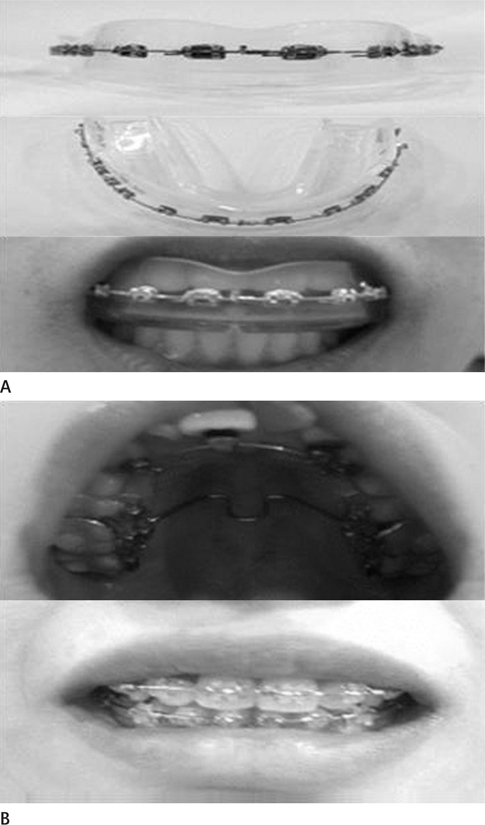
Fig. 1 Photograph of home-made orthodontic device model in group A (A) and patient wearing therapeutic orthodontic device in group B (B).
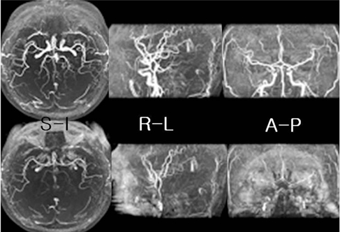
Fig. 2 Three dimensional time of flight magnetic resonance angiographys in control (group A) before (top images) and after wearing the orthodontic device (bottom images). Maximal intensity projection images of various directions without wearing the device show well delineation of distal cerebral arteries. However, after wearing the device, there are multiple arteries with signal loss and poor margination. Note.-S-I = superior-inferior direction, R-L = right-left direction, A-P = anterior-posterior direction

Fig. 3 Three dimensional phase contrast magnetic resonance angiographys in control (group A) before (top images) and after wearing the orthodontic device (bottom images). All maximal intensity projection images show well marginated cerebral arteries regardless of wearing the device. Venous flows are detected in both transverse and sigmoid sinuses. Note.-S-I = superior-inferior direction, R-L = right-left direction, A-P = anterior-posterior direction
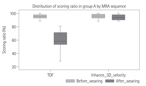
Fig. 4 Distribution of scoring ratio in controls (group A). Scoring ratios (percentage) measured from 3D TOF method show significant difference between group before wearing device and group after wearing device. In 3D PC method, there are no differences of scoring ratios in two groups. 'Inhance 3D velocity' is MRA means 3D PC technique. Note.-MRA = magnetic resonance angiography, PC = phase contrast, TOF = time of flight, 3D = 3 dimensional
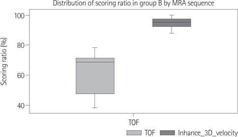
Fig. 5 Distribution of scoring ratio in patients (group B). In patients wearing actual orthodontic devices, Difference of scoring ratios between group with 3D TOF method (average 68%) and group with 3D PC method (average 94%) is significant. 'Inhance 3D velocity' means MRA using 3D PC technique. Note.-MRA = magnetic resonance angiography, PC = phase contrast, TOF = time of flight, 3D = 3 dimensional
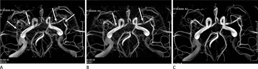
Fig. 6 Comparison of 3D PC MRAs by variable velocity encoding (VENC) values (25 cm/sec in A, 35 cm/sec in B, 45 cm/sec in C). MRAs scanned by 25 and 35 cm/sec VENC values show multifocal signal losses mimicking stenosis or slow flow in cerebral arteries (arrows). However, these phenomena disappear in C (45 cm/sec VENC value). Note.-3D PC MRA = 3 dimensional phase contrast magnetic resonance angiography
Reference
-
1. Acheson J, Boyd WN, Hugh AE, Hutchinson EC. Cerebral angiography in ischemic cerebrovascular disease. Arch Neurol. 1969; 20:527–532.2. White PM, Wardlaw JM, Easton V. Can noninvasive imaging accurately depict intracranial aneurysms? A systematic review. Radiology. 2000; 217:361–337.3. Yang JJ, Hill MD, Morrish WF, Hudon ME, Barber PA, Demchuk AM, et al. Comparison of pre- and postcontrast 3D time-of-flight MR angiography for the evaluation of distal intracranial branch occlusions in acute ischemic stroke. AJNR Am J Neuroradiol. 2002; 23:557–567.4. Remonda L, Senn P, Barth A, Arnold M, Lövblad KO, Schroth G. Contrast-enhanced 3D MR angiography of the carotid artery: comparison with conventional digital subtraction angiography. AJNR Am J Neuroradiol. 2002; 23:213–219.5. Campeau NG, Huston J 3rd, Bernstein MA, Lin C, Gibbs GF. Magnetic resonance angiography at 3.0 Tesla: initial clinical experience. Top Magn Reson Imaging. 2001; 12:183–120.6. Al-Kwifi O, Emery DJ, Wilman AH. Vessel contrast at three Tesla in time-of-flight magnetic resonance angiography of the intracranial and carotid arteries. Magn Reson Imaging. 2002; 20:181–187.7. Frayne R, Goodyear BG, Dickhoff P, Lauzon ML, Sevick RJ. Magnetic resonance imaging at 3.0 Tesla: challenges and advantages in clinical neurological imaging. Invest Radiol. 2003; 38:385–340.8. Wilms G, Bosmans H, Demaerel P, Marchal G. Magnetic resonance angiography of the intracranial vessels. Eur J Radiol. 2001; 38:10–18.9. Walker MT, Tsai J, Parish T, Tzung B, Shaibani A, Krupinski E, et al. MR angiographic evaluation of platinum coil packs at 1.5T and 3T: an in vitro assessment of artifact production: technical note. AJNR Am J Neuroradiol. 2005; 26:848–885.10. Willinek WA, Born M, Simon B, Tschampa HJ, Krautmacher C, Gieseke J, et al. Time-of-flight MR angiography: comparison of 3.0-T imaging and 1.5-T imaging--initial experience. Radiology. 2003; 229:913–920.11. Yu HS, Ryu YK, Lee JY. A study on the distributions and trends in malocclusion patients from department of orthodontics, college of dentistry, Yonsei university. Korean J Orthod. 1999; 29:267–276.12. Lee YK, Cha JY, Yu HS, Hwang CJ. Effect of metal primer and thermocycling on shear bonding strength between the orthodontic bracket and gold alloy. Korean J Orthod. 2009; 39:320–329.13. Liu JK, Chang LT, Chuang SF, Shieh DB. Shear bond strengths of plastic brackets with a mechanical base. Angle Orthod. 2002; 72:141–145.14. Gizewski ER, Ladd ME, Paul A, Wanke I, Göricke S, Forsting M. Water excitation: a possible pitfall in cerebral time-of-flight angiography. AJNR Am J Neuroradiol. 2005; 26:152–155.15. Griswold MA, Jakob PM, Nittka M, Goldfarb JW, Haase A. Partially parallel imaging with localized sensitivities (PILS). Magn Reson Med. 2000; 44:602–609.16. Weiger M, Pruessmann KP, Leussler C, Röschmann P, Boesiger P. Specific coil design for SENSE: a six-element cardiac array. Magn Reson Med. 2001; 45:495–504.17. Wilson GJ, Hoogeveen RM, Willinek WA, Muthupillai R, Maki JH. Parallel imaging in MR angiography. Top Magn Reson Imaging. 2004; 15:169–185.18. Hiai Y, Kakeda S, Sato T, Ohnari N, Moriya J, Kitajima M, et al. 3D TOF MRA of intracranial aneurysms at 1.5 T and 3 T: influence of matrix, parallel imaging, and acquisition time on image quality - a vascular phantom study. Acad Radiol. 2008; 15:635–664.19. Marks MP, Pelc NJ, Ross MR, Enzmann DR. Determination of cerebral blood flow with a phase-contrast cine MR imaging technique: evaluation of normal subjects and patients with arteriovenous malformations. Radiology. 1992; 182:467–476.20. Oelerich M, Lentschig MG, Zunker P, Reimer P, Rummeny EJ, Schuierer G. Intracranial vascular stenosis and occlusion: comparison of 3D time-of-flight and 3D phase-contrast MR angiography. Neuroradiology. 1998; 40:567–573.21. Shonai T, Carpenter JS, Lemieux SK, Harada K, Omori K, Kaneko N, et al. Improvement of vessel visibility in time-of-flight MR angiography of the brain. J Magn Reson Imaging. 2008; 27:1362–1370.22. Grayev A, Shimakawa A, Cousins J, Turski P, Brittain J, Reeder S. Improved time-of-flight magnetic resonance angiography with IDEAL water-fat separation. J Magn Reson Imaging. 2009; 29:1367–1374.23. Dion JE, Gates PC, Fox AJ, Barnett HJ, Blom RJ. Clinical events following neuroangiography: a prospective study. Stroke. 1987; 18:997–1004.24. Grzyska U, Freitag J, Zeumer H. Selective cerebral intraarterial DSA. Complication rate and control of risk factors. Neuroradiology. 1990; 32:296–229.25. Randoux B, Marro B, Koskas F, Duyme M, Sahel M, Zouaoui A, et al. Carotid artery stenosis: prospective comparison of CT, three-dimensional gadolinium-enhanced MR, and conventional angiography. Radiology. 2001; 220:179–185.26. Hirai T, Korogi Y, Ono K, Nagano M, Maruoka K, Uemura S, et al. Prospective evaluation of suspected stenoocclusive disease of the intracranial artery: combined MR angiography and CT angiography compared with digital subtraction angiography. AJNR Am J Neuroradiol. 2002; 23:93–101.27. Bosmans H, Marchal G, Lukito G, Yicheng N, Wilms G, Laub G, et al. Time-of-flight MR angiography of the brain: comparison of acquisition techniques in healthy volunteers. AJR Am J Roentgenol. 1995; 164:161–167.28. White PM, Teasdale EM, Wardlaw JM, Easton V. Intracranial aneurysms: CT angiography and MR angiography for detection prospective blinded comparison in a large patient cohort. Radiology. 2001; 219:739–749.29. Ozsarlak O, Van Goethem JW, Parizel PM. 3D time-of-flight MR angiography of the intracranial vessels: optimization of the technique with water excitation, parallel acquisition, eight-channel phased-array head coil and low-dose contrast administration. Eur Radiol. 2004; 14:2067–2071.30. Korosec FR, Mistretta CA. MR angiography: basic principles and theory. Magn Reson Imaging Clin N Am. 1998; 6:223–256.31. Hollnagel DI, Summers PE, Poulikakos D, Kollias SS. Comparative velocity investigations in cerebral arteries and aneurysms: 3D phase-contrast MR angiography, laser Doppler velocimetry and computational fluid dynamics. NMR Biomed. 2009; 22:795–808.
- Full Text Links
-
- Actions
-
Cited
- CITED
-
- Close
- Share
-
- Similar articles
-
- Artifacts in MR Angiography of the Intracranial Vessels Using the 3D TOF and 3D PC Techniques
- Accelerated Time-of-Flight Magnetic Resonance Angiography with Sparse Undersampling and Iterative Reconstruction for the Evaluation of Intracranial Arteries
- Two-Dimensional Image-Based Respiratory Navigator for Free-Breathing Coronary Magnetic Resonance Angiography
- Development and Feasibility Study for Phase Contrast MR Angiography at Low Tesla Open-MRI System
- Detection of Intracranial Aneurysms by MRA and Conventional Angiography
