Intrahepatic Extramedullary Hematopoiesis Mimicking a Hypervascular Hepatic Neoplasm on Dynamic- and SPIO-Enhanced MRI
- Affiliations
-
- 1Department of Radiology, Seoul National University Hospital, Seoul, Korea. shkim@radcom.snu.ac.kr
- 2Institute of Radiation Medicine, Seoul National University Hospital, Seoul, Korea.
- KMID: 1100101
- DOI: http://doi.org/10.3348/kjr.2008.9.s.s34
Abstract
- We present a rare case of a focal intrahepatic extramedullary hematopoiesis (EMH) that mimicked a hypervascular hepatic neoplasm in a 33-year-old woman with idiopathic myelofibrosis. The lesion showed homogeneous and persistent enhancement on both contrast-enhanced CT and gadolinium-enhanced dynamic MR imaging. The lesion did not demonstrate an apparent signal drop on a T2*-weighted sequence following administration of a superparamagnetic iron-oxide agent (SHU 555A). A hepatocellular adenoma was the initial radiological diagnosis. To the best of our knowledge, this is the first report of a histopathologically proven intrahepatic EMH evaluated with dynamic- and SPIO-enhanced MRI.
Keyword
MeSH Terms
Figure
-
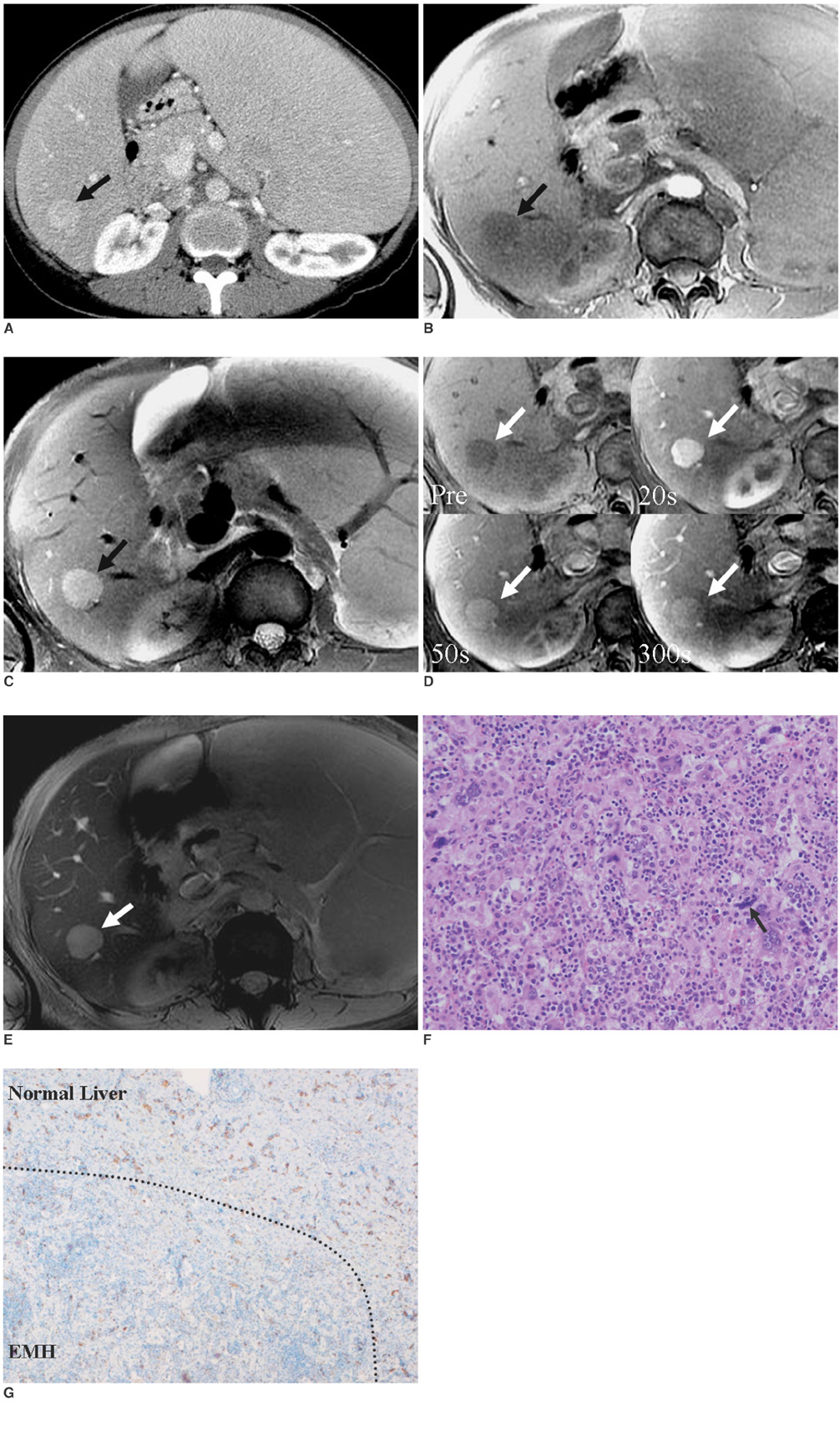
Fig. 1 Intrahepatic extramedullary hematopoiesis in 33-year-old woman. A. 33-year-old woman with history of idiopathic myelofibrosis. On contrast-enhanced CT obtained in equilibrium phase, 2-cm-sized, well-defined and homogeneously enhancing mass (arrow) is seen in segment VI of liver. Liver and spleen are diffusely enlarged. B. Lesion (arrow) shows homogeneous low signal intensity on in-phase T1-weighted image (repetition time [TR]/echo time [TE] = 140 msec/2.4 msec, flip angle [FA] = 70°). C. Lesion (arrow) shows high signal intensity on fat-saturated, T2-weighted fast spin echo image (TR/TE = 12,857.1/100.7). D. On serial gadolinium-BOPTA dynamic MR scans, lesion (arrows) shows homogeneous and intense enhancement on arterial phase (20 seconds after gadolinium administration, upper right) compared with pre-contrast T1-weighted image (TR/TE = 4.7/2.2, FA = 10°) (upper left); it enhances persistently through portal (50 seconds, lower left) to delayed phases (5 minutes, lower right). E. On SPIO-enhanced, T2*-weighted gradient echo image (TR/TE = 130.0/9.7, FA = 30°) obtained 10 minutes after SPIO administration, there is no signal drop in lesion (arrow). F. On the photomicrograph (Hematoxylin & Eosin staining; ×200), megakaryocytes (arrow) and pleomorphic groups of erythroid and myeloid precursors that represent three hematologic precursor cell-lines, are seen insinuating hepatocyte cords. G. Immunohistochemical staining using CD68 marker (×40), reveals that cells in extramedullary hematopoiesis are positive for CD 68 (stained with brown color), but degree of staining in lesion (lower part of dotted line) is much weaker than that in normal liver (upper part of dotted line).
Reference
-
1. Aytac S, Fitoz S, Akyar S, Atasoy C, Erekul S. Focal intrahepatic extramedullary hematopoiesis: color Doppler US and CT findings. Abdom Imaging. 1999. 24:366–368.2. Bradley MJ, Metreweli C. Sonography of extramedullary hematopoiesis of the liver. AJR Am J Roentgenol. 1990. 154:900–901.3. Dardi LE, Marzano M, Froula E. Fine needle aspiration cytologic diagnosis of focal intrahepatic extramedullary hematopoiesis. Acta Cytol. 1990. 34:567–569.4. Dewar G, Leung NW, Ng HK, Bradley M, Li AK. Massive, solitary, intrahepatic, extramedullary hematopoietic tumor in thalassemia. Surgery. 1990. 107:704–707.5. Warshauer DM, Schiebler ML. Intrahepatic extramedullary hematopoiesis: MR, CT, and sonographic appearance. J Comput Assist Tomogr. 1991. 15:683–685.6. Wong Y, Chen F, Tai KS, Yip LK, Tsang KW, Chan FL, et al. Imaging features of focal intrahepatic extramedullary haematopoiesis. Br J Radiol. 1999. 72:906–910.7. Kwak HS, Lee JM. CT findings of extramedullary hematopoiesis in the thorax, liver and kidneys, in a patient with idiopathic myelofibrosis. J Korean Med Sci. 2000. 15:460–462.8. Navarro M, Crespo C, Perez L, Martinez C, Galant J, Gonzalez I. Massive intrahepatic extramedullary hematopoiesis in myelofibrosis. Abdom Imaging. 2000. 25:184–186.9. Gupta P, Naran A, Auh YH, Chung JS. Focal intrahepatic extramedullary hematopoiesis presenting as fatty lesions. AJR Am J Roentgenol. 2004. 182:1031–1032.10. Jelali MA, Luciani A, Kobeiter H, Zafrani S, Anglade MC, Zegai B, et al. MRI features of intrahepatic extramedullary haematopoiesis in sickle cell anaemia. Cancer Imaging. 2006. 6:182–185.11. Semelka RC, Martin DR, Balci C, Lance T. Focal liver lesions: comparison of dual-phase CT and multisequence multiplanar MR imaging including dynamic gadolinium enhancement. J Magn Reson Imaging. 2001. 13:397–401.12. Reimer P, Jahnke N, Fiebich M, Schima W, Deckers F, Marx C, et al. Hepatic lesion detection and characterization: value of nonenhanced MR imaging, superparamagnetic iron oxide-enhanced MR imaging, and spiral CT-ROC analysis. Radiology. 2000. 217:152–158.13. Kim SH, Lee JM, Han JK, Lee JY, Kang WJ, Jang JY, et al. MDCT and superparamagnetic iron oxide (SPIO)-enhanced MR findings of intrapancreatic accessory spleen in seven patients. Eur Radiol. 2006. 16:1887–1897.14. Wang YX, Hussain SM, Krestin GP. Superparamagnetic iron oxide contrast agents: physicochemical characteristics and applications in MR imaging. Eur Radiol. 2001. 11:2319–2331.15. Fukuda Y, Ando K, Ishikura R, Kotoura N, Tsuda N, Kato N, et al. Superparamagnetic iron oxide (SPIO) MRI contrast agent for bone marrow imaging: differentiating bone metastasis and osteomyelitis. Magn Reson Med Sci. 2006. 5:191–196.16. Weissleder R, Elizondo G, Wittenberg J, Rabito CA, Bengele HH, Josephson L. Ultrasmall superparamagnetic iron oxide: characterization of a new class of contrast agents for MR imaging. Radiology. 1990. 175:489–493.17. Wiener MD, Halvorsen RA Jr, Vollmer RT, Foster WL, Roberts L Jr. Focal intrahepatic extramedullary hematopoiesis mimicking neoplasm. AJR Am J Roentgenol. 1987. 149:1171–1172.18. Abbitt PL, Teates CD. The sonographic appearance of extramedullary hematopoiesis in the liver. J Clin Ultrasound. 1989. 17:280–282.
- Full Text Links
-
- Actions
-
Cited
- CITED
-
- Close
- Share
-
- Similar articles
-
- Periportal Extramedullary Hematopoiesis
- Saccular Aneurysm of Intraheparic Portal Vein Mimicking Hypervascular Hepatic Mass: A Case Report
- Detectability of Hepatocellular Carcinoma: Comparison of Gd-DT PA-Enhanced and SPIO-Enhanced MR Imaging
- A Case of Extramedullary Hematopoiesis Associated with Congenital Dyserythropoietic Anemia
- Cutaneous Extramedullary Hematopoiesis in Idiopathic Myelofibrosis
