Korean J Radiol.
2008 Apr;9(2):102-110. 10.3348/kjr.2008.9.2.102.
Estimating the Absorbed Dose to Critical Organs During Dual X-ray Absorptiometry
- Affiliations
-
- 1Medical Physics Department, Tarbiat Modares University, Tehran, Iran. mokhtarm@modares.ac.ir
- 2Medical Physics Department, Iran University of Medical Sciences, Tehran, Iran.
- 3Metabolism and Endocrine Research Center, Tehran Medical Sciences University, Tehran, Iran.
- 4Medical Radiation Department, Azad University, Tehran, Iran.
- KMID: 1098188
- DOI: http://doi.org/10.3348/kjr.2008.9.2.102
Abstract
OBJECTIVE
The purpose of this study is to estimate a patient's organ dose (effective dose) during performance of dual X-ray absorptiometry by using the correlations derived from the surface dose and the depth doses in an anthropomorphic phantom. MATERIALS AND METHODS: An anthropomorphic phantom was designed and TLDs (Thermoluminescent Dosimeters) were placed at the surface and these were also inserted at different depths of the thyroid and uterus of the anthropomorphic phantom. The absorbed doses were measured on the phantom for the spine and femur scan modes. The correlation coefficients and regression functions between the absorbed surface dose and the depth dose were determined. The derived correlation was then applied for 40 women patients to estimate the depth doses to the thyroid and uterus. RESULTS: There was a correlation between the surface dose and depth dose of the thyroid and uterus in both scan modes. For the women's dosimetry, the average surface doses of the thyroid and uterus were 1.88 (micro)Gy and 1.81 (micro)Gy, respectively. Also, the scan center dose in the women was 5.70 (micro)Gy. There was correlation between the thyroid and uterus surface doses, and the scan center dose. CONCLUSION: We concluded that the effective dose to the patient's critical organs during dual X-ray absorptiometry can be estimated by the correlation derived from phantom dosimetry.
MeSH Terms
Figure
-
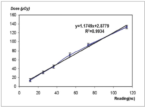
Fig. 1 Calibration curve of TLDs.
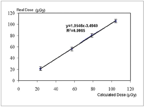
Fig. 2 Correction factor curve for TLDs.
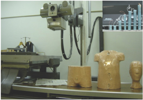
Fig. 3 Front view of anthropomorphic phantom with depth probes.
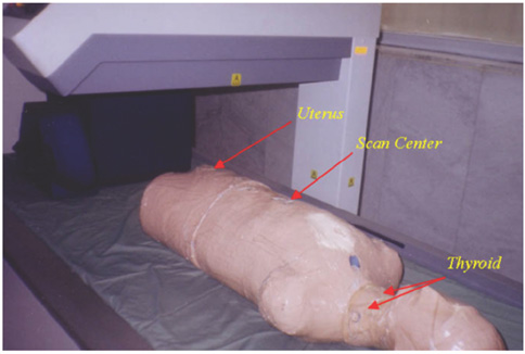
Fig. 4 Anthropomorphic phantom in measurement position.
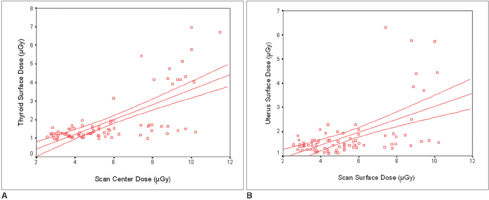
Fig. 5 Scatter plot between centers of scan dose with thyroid surface dose (A) and uterus surface dose (µGy) (B) with 95% confidence interval.
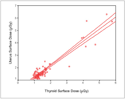
Fig. 6 Scatter plot between uterus surface dose and thyroid surface dose (µGy) with 95% confidence interval.
Reference
-
1. Blinov NN, Gubenko MB, Utkin PM. Development of osteodensitometric equipment. Biomed Eng. 2002. 36:36–40.2. Diez F. Guidelines for the diagnosis of osteoporosis by densitometric methods. J Manipulative Physiol Ther. 2002. 25:403–415.3. Boudousq V, Kotzki PO, Dinten JM, Barrau C, Robert-Coutant C, Thomas E, et al. Total dose incurred by patients and staff from BMD measurement using a new 2D digital bone densitometer. Osteoporos Int. 2003. 14:263–269.4. Steel SA, Baker AJ, Saunderson JR. An assessment of the radiation dose to patients and staff from a Lunar Expert-XL fan beam. Physiol Meas. 1998. 19:17–26.5. Eiken P, Barenholdt O, Jensen LB, Gram J, Nielson SP. Switching from DXA pencil beam to fan beam. I: studies in vitro at four centers. Bone. 1994. 15:667–670.6. Eiken P, Kolthoff N, Barenholdt O, Hermansen H, Nielson SP. Switching from DXA pencil beam to fan beam. II: studies in vivo. Bone. 1994. 15:671–676.7. Royal college of radiologist and national radiological protection board. Patient dose reduction in diagnostic radiology. 1991. 1. London: HMSO;3. (documents of the NRPB).8. Popovic M, McNiell FE, Webber CE, Chettle DR. The effect of lead in bone densitometry. Nuclear Instruments and Methods in Physics Research Section B. 2004. 213:599–602.9. Culton N, Pocock N. Evaluation of three spine phantoms for DXA QC. Bone. 2000. 27:48S.10. Dequeker J, Pearson J, Reeve J, Henley M, Bright J, Felsenberg D, et al. Dual x-ray absorptiometry--cross-calibration and normative reference ranges for the spin: results of a European Community Concerted Action. Bone. 1995. 17:247–254.11. Kalender WA, Felsenberg D, Genant HK, Fischer M, Dequeker J, Reeve J. The European Spine Phantom-a tool for standardization and quality control in spinal bone mineral measurements by DXA and QCT. Eur J Radiol. 1995. 20:83–92.12. Abrahamsen B, Gram J, Hansen TB, Beck-Nielsen H. Cross calibration of QDR-2000 and QDR-1000 dual energy X-ray densitometers for bone mineral and soft-tissue measurements. Bone. 1995. 16:385–390.13. Njeh CF, Samat SB, Nightingale A, McNeil EA, Boivin CM. Radiation dose and in vitro precision in paediatric bone mineral density measurement using dual X-ray absorptiometry. Br J Radiol. 1997. 70:719–727.14. Njeh CF, Apple K, Temperton DH, Boivin CM. Radiological assessment of a new bone densitometer-the Lunar EXPERT. Br J Radiol. 1996. 69:335–340.15. Blake GM, Patel R, Lewis MK, Batchelor S. New generation dual X-ray absorptiometry: a comparison of pencil beam and fan beam systems. Br J Radiol. 1996. 11:S157.16. Damilakis J, Perisinakis K, Vrahoriti H, Kontakis G, Varveris H, Gourtsoyiannis N. Embryo/fetus radiation dose and risk from dual X-ray absorptiometry examinations. Osteoporos Int. 2002. 13:716–722.17. Buroker KD. DPX series operator's manual. 1998. 45–80.18. Hughes JS, Shaw KB, O'Riordan MC. Radiation exposure of the UK population: 1988 review. 1989. London: HMSO;32–36. National Radiation Protection Board (Report NRPB-R227).19. Boutros M. Radiation dose assessment to patients and staff from the new LEXXOS bone dosimeter MSc thesis in Medical Engineering and Physics. 2003. King's collage London;45–63.20. Koo WW, Walters J, Bush AJ. Technical considerations of dual-energy X-ray absorptiometry-based bone mineral measurements for pediatric studies. J Bone Miner Res. 1995. 10:1998–2004.21. Hart D, Jones DG, Wall BF. Coefficient for estimating effective doses from paediatric x-ray examinations. 1996. London: HMSO;24–27. (NRPB-R279).
- Full Text Links
-
- Actions
-
Cited
- CITED
-
- Close
- Share
-
- Similar articles
-
- Measurement and Interpretation of Dual-Energy X-ray Absorptiometry Bone Density Measurements
- Absorbed and effective dose for periapical radiography using portable and wall type dental X-ray machines
- Reliability of Singh's index Checked by the Dual Photon X-ray Absorptiometry(LUNAR D.P.X)
- Radiation Dose Calculation in the Surrounding Organs during Brachytherapy of Prostate Cancer
- Absorbed doses in organs of the head and neck from conventional temporomandibular joint tomography
