Korean J Radiol.
2008 Apr;9(2):95-101. 10.3348/kjr.2008.9.2.95.
Ultra-Low-Dose MDCT of the Chest: Influence on Automated Lung Nodule Detection
- Affiliations
-
- 1Department of Radiology and Center for Imaging Science, Samsung Medical Center, Sungkyunkwan University School of Medicine, Seoul, Korea. mj1.chung@samsung.com
- KMID: 1098187
- DOI: http://doi.org/10.3348/kjr.2008.9.2.95
Abstract
OBJECTIVE
To evaluate the relationship between CT dose and the performance of a computer-aided diagnosis (CAD) system, and to determine how best to minimize patient exposure to ionizing radiation while maintaining sufficient image quality for automated lung nodule detection, by the use of lung cancer screening CT. MATERIALS AND METHODS: Twenty-five asymptomatic volunteers participated in the study. Each volunteer underwent a low-dose CT scan without contrast enhancement (multidetector CT with 16 detector rows, 1.25 mm section thickness, 120 kVp, beam pitch 1.35, 0.6 second rotation time, with 1.25 mm thickness reconstruction at 1.25 mm intervals) using four different amperages 32, 16, 8, and 4 mAs. All series were analyzed using a commercially available CAD system for automatic lung nodule detection and the results were reviewed by a consensus reading by two radiologists. The McNemar test and Kappa analysis were used to compare differences in terms of the abilities to detect pulmonary nodules. RESULTS: A total of 78 non-calcified true nodules were visualized in the 25 study subjects. The sensitivities for nodule detection were as follows: 72% at 32 mAs, 64% at 16 mAs, 59% at 8 mAs, and 40% at 4 mAs. Although the overall nodule-detecting performance was best at 32 mAs, no significant difference in nodule detectability was observed between scans at 16 mAs or 8 mAs versus 32 mAs. However, scans performed at 4 mAs were significantly inferior to those performed at 32 mAs (p < 0.001). CONCLUSION: Reducing the radiation dose (i.e. reducing the amperage) lowers lung nodule detectability by CAD. However, relatively low dose scans were found to be acceptable and to cause no significant reduction in nodule detectability versus usual low-dose CT.
MeSH Terms
Figure
-
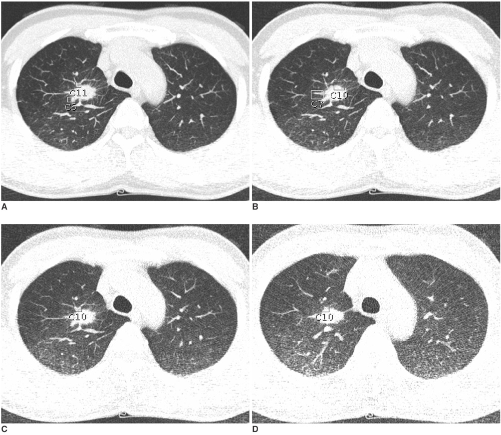
Fig. 1 Transverse CT scans were acquired at tube currents of 32 (A), 16 (B), 8 (C), and 4 (D) mAs. Note that image quality was reduced and noise was increased as tube currents were decreased.

Fig. 2 Five-mm nodule in right lower lobe superior segment, which was detected at 32 (A), 16 and 8 mAs/slice (not shown in this figure), but not at 4 mAs/slice (C) on the CAD system. Note that volume-rendering image obtained at 32 mAs (B) is sharper than that obtained at 4 mAs (D).
Reference
-
1. Chong S, Lee KS, Chung MJ, Kim TS, Kim H, Kwon OJ, et al. Lung cancer screening with low-dose helical CT in Korea: experiences at the Samsung Medical Center. J Korean Med Sci. 2005. 20:402–408.2. Gergely I, Neumann C, Reiger F, Dorffner R. Lung nodule detection with ultra-low-dose CT in routine follow-up of cancer patients. Rofo. 2005. 177:1077–1083.3. Karabulut N, Toru M, Gelebek V, Gulsun M, Ariyurek OM. Comparison of low-dose and standard-dose helical CT in the evaluation of pulmonary nodules. Eur Radiol. 2002. 12:2764–2769.4. Nitta N, Takahashi M, Murata K, Morita R. Ultra low-dose helical CT of the chest: evaluation in clinical cases. Radiat Med. 1999. 17:1–7.5. Nitta N, Takahashi M, Murata K, Morita R. Ultra low-dose helical CT of the chest. AJR Am J Roentgenol. 1998. 171:383–385.6. Kaneko M, Eguchi K, Ohmatsu H, Kakinuma R, Naruke T, Suemasu K, et al. Peripheral lung cancer: screening and detection with low-dose spiral CT versus radiography. Radiology. 1996. 201:798–802.7. Marten K, Engelke C, Seyfarth T, Grillhosl A, Obenauer S, Rummeny EJ. Computer-aided detection of pulmonary nodules: influence of nodule characteristics on detection performance. Clin Radiol. 2005. 60:196–206.8. Armato SG 3rd, Roy AS, Macmahon H, Li F, Doi K, Sone S, et al. Evaluation of automated lung nodule detection on low-dose computed tomography scans from a lung cancer screening program(1). Acad Radiol. 2005. 12:337–346.9. Lee JW, Goo JM, Lee HJ, Kim JH, Kim S, Kim YT. The potential contribution of a computer-aided detection system for lung nodule detection in multidetector row computed tomography. Invest Radiol. 2004. 39:649–655.10. Awai K, Murao K, Ozawa A, Komi M, Hayakawa H, Hori S, et al. Pulmonary nodules at chest CT: effect of computer-aided diagnosis on radiologists' detection performance. Radiology. 2004. 230:347–352.11. Arimura H, Katsuragawa S, Suzuki K, Li F, Shiraishi J, Sone S, et al. Computerized scheme for automated detection of lung nodules in low-dose computed tomography images for lung cancer screening. Acad Radiol. 2004. 11:617–629.12. Goo JM, Lee JW, Lee HJ, Kim S, Kim JH, Im JG. Automated lung nodule detection at low-dose CT: preliminary experience. Korean J Radiol. 2003. 4:211–216.13. Naidich DP, Marshall CH, Gribbin C, Arams RS, McCauley DI. Low-dose CT of the lungs: preliminary observations. Radiology. 1990. 175:729–731.14. Flehinger BJ, Melamed MR. Current status of screening for lung cancer. Chest Surg Clin N Am. 1994. 4:1–15.15. Flehinger BJ, Kimmel M, Melamed MR. The effect of surgical treatment on survival from early lung cancer. Implications for screening. Chest. 1992. 101:1013–1018.16. Swensen SJ, Jett JR, Hartman TE, Midthun DE, Sloan JA, Sykes AM, et al. Lung cancer screening with CT: Mayo Clinic experience. Radiology. 2003. 226:756–761.17. Henschke CI. Early lung cancer action project: overall design and findings from baseline screening. Cancer. 2000. 89:2474–2482.18. Nickoloff EL, Alderson PO. Radiation exposures to patients from CT: reality, public perception, and policy. AJR Am J Roentgenol. 2001. 177:285–287.19. Remy-Jardin M, Remy J. Spiral CT angiography of the pulmonary circulation. Radiology. 1999. 212:615–636.20. Zhu X, Yu J, Huang Z. Low-dose chest CT: optimizing radiation protection for patients. AJR Am J Roentgenol. 2004. 183:809–816.21. Jung KJ, Lee KS, Kim SY, Kim TS, Pyeun YS, Lee JY. Low-dose, volumetric helical CT: image quality, radiation dose, and usefulness for evaluation of bronchiectasis. Invest Radiol. 2000. 35:557–563.22. Brenner DJ. Radiation risks potentially associated with low-dose CT screening of adult smokers for lung cancer. Radiology. 2004. 231:440–445.23. Rusinek H, Naidich DP, McGuinness G, Leitman BS, McCauley DI, Krinsky GA, et al. Pulmonary nodule detection: low-dose versus conventional CT. Radiology. 1998. 209:243–249.24. Mayo JR, Kim KI, MacDonald SL, Johkoh T, Kavanagh P, Coxson HO, et al. Reduced radiation dose helical chest CT: effect on reader evaluation of structures and lung findings. Radiology. 2004. 232:749–756.
- Full Text Links
-
- Actions
-
Cited
- CITED
-
- Close
- Share
-
- Similar articles
-
- Automated Lung Nodule Detection at Low-Dose CT: Preliminary Experience
- Screening for Lung Cancer
- Lung Cancer Screening with Low-dose Computed Tomography
- Detection of Pulmonary Metastatic Nodules: Usefulness of Low-dose Multidetector CT in Comparison with Chest Radiograph
- Lung cancer screening with low-dose chest computed tomography: recent radiologic advances
