Korean J Radiol.
2003 Dec;4(4):211-216. 10.3348/kjr.2003.4.4.211.
Automated Lung Nodule Detection at Low-Dose CT: Preliminary Experience
- Affiliations
-
- 1Department of Radiology, Seoul National University College of Medicine, and the Institute of Radiation Medicine, SNUMRC. jmgoo@plaza.snu.ac.kr
- 2Human Information Processing Team, Electronics and Telecommunications Research Institute
- KMID: 1111249
- DOI: http://doi.org/10.3348/kjr.2003.4.4.211
Abstract
OBJECTIVE
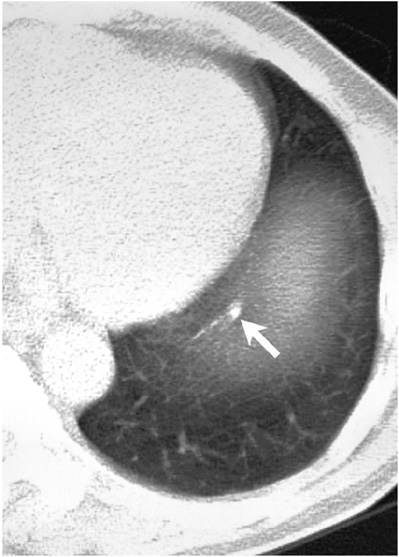
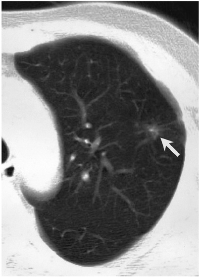
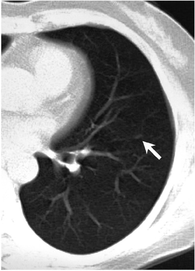
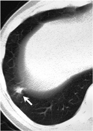
To determine the usefulness of a computer-aided diagnosis (CAD) system for the automated detection of lung nodules at low-dose CT. MATERIALS AND METHODS : A CAD system developed for detecting lung nodules was used to process the data provided by 50 consecutive low-dose CT scans. The results of an initial report, a second look review by two chest radiologists, and those obtained by the CAD system were compared, and by reviewing all of these, a gold standard was established. RESULTS : By applying the gold standard, a total of 52 nodules were identified (26 with a diameter < or =5 mm; 26 with a diameter > 5 mm). Compared to an initial report, four additional nodules were detected by the CAD system. Three of these, identified only at CAD, formed part of the data used to derive the gold standard. For the detection of nodules > 5 mm in diameter, sensitivity was 77% for the initial report, 88% for the second look review, and 65% for the CAD system. There were 8.0+/-5.2 false-positive CAD results per CT study. CONCLUSION : These preliminary results indicate that a CAD system may improve the detection of pulmonary nodules at low-dose CT.
Keyword
MeSH Terms
Figure
Reference
-
1. Henschke CI, McCauley DI, Yankelevitz DF, et al. Early Lung Cancer Action Project: overall design and findings from baseline screening. Lancet. 1999. 354:99–105.2. Diederich S, Wormanns D, Semik M, et al. Screening for early lung cancer with low-dose spiral CT: prevalence in 817 asymptomatic smokers. Radiology. 2002. 222:773–781.3. Swensen SJ, Jett JR, Sloan JA, et al. Screening for lung cancer with low-dose spiral computed tomography. Am J Respir Crit Care Med. 2002. 165:508–513.4. Kakinuma R, Ohmatsu H, Kaneko M, et al. Detection failures in spiral CT screening for lung cancer: analysis of CT findings. Radiology. 1999. 212:61–66.5. Li F, Sone S, Abe H, MacMahon H, Armato SG III, Doi K. Lung cancers missed at low-dose helical CT screening in a general population: comparison of clinical, histopathologic, and imaging findings. Radiology. 2002. 225:673–683.6. Giger ML, Karssemeijer N, Armato SG III. Computer-aided diagnosis in medical imaging. IEEE Trans Med Imaging. 2001. 20:1205–1208.7. Giger ML, Bae KT, MacMahon H. Computerized detection of pulmonary nodules in computed tomography images. Invest Radiol. 1994. 29:459–465.8. Kanazawa K, Kawata Y, Niki N, et al. Computer-aided diagnosis for pulmonary nodules based on helical CT images. Comput Med Imaging Graph. 1998. 22:157–167.9. Armato SG III, Giger ML, MacMahon H. Automated detection of lung nodules on CT scans: preliminary results. Med Phys. 2001. 28:1552–1561.10. Ko JP, Betke M. Chest CT: automated nodule detection and assessment of change over time-preliminary experience. Radiology. 2001. 218:267–273.11. Lee Y, Hara T, Fujita H, et al. Automated detection of pulmonary nodules in helical CT images based on an improved template-matching technique. IEEE Trans Med Imaging. 2001. 20:595–604.12. Gurcan MN, Sahiner B, Petrick N, et al. Lung nodule detection on thoracic computed tomography images: preliminary evaluation of a computer-aided diagnosis system. Med Phys. 2002. 29:2552–2558.13. Wormanns D, Fiebich M, Saidi M, Diederich S, Heindel W. Automatic detection of pulmonary nodules at spiral CT: clinical application of a computer-aided diagnosis system. Eur Radiol. 2002. 12:1052–1057.14. Brown MS, Goldin JG, Suh RD, McNitt-Gray MF, Sayre JW, Aberle DR. Lung micronodules: automated method for detection at thin-section CT: initial experience. Radiology. 2003. 226:256–262.15. Armato SG III, Li F, Giger ML, MacMahon H, Sone S, Doi K. Lung cancer: performance of automated lung nodule detection applied to cancers missed in a CT screening program. Radiology. 2002. 225:685–692.16. Wormanns D, Diederich S, Lentschig MG, Winter F, Heindel W. Spiral CT of pulmonary nodules: interobserver variation in assessment of lesion size. Eur Radiol. 2000. 10:710–713.
- Full Text Links
- Actions
-
Cited
- CITED
-
- Close
- Share
- Similar articles
-
- Ultra-Low-Dose MDCT of the Chest: Influence on Automated Lung Nodule Detection
- Screening for Lung Cancer
- Lung Cancer Screening with Low-Dose Chest CT: Current Issues
- Characteristics of Pulmonary Nodules in Current Smoker
- Studies and Real-World Experience Regarding the Clinical Application of Artificial Intelligence Software for Lung Nodule Detection


