A Case of Multiple Mucosa-Associated Lymphoid Tissue (MALT) Lymphoma of the Colon Identified as Simple Mucosal Discoloration
- Affiliations
-
- 1Department of Internal Medicine, Seonam University School of Medicine, Korea. lsmed@hanmail.net
- 2Department of Anatomic Pathology, Seonam University School of Medicine, Korea.
- 3Miraero 21 Hospital, Gwangju, Korea.
- KMID: 1095213
- DOI: http://doi.org/10.3346/jkms.2005.20.2.325
Abstract
- Most colonic multiple mucosa-associated lymphoid tissue (MALT) lymphomas are confirmed with a histologic and immunohistochemical staining of the mucosal biopsy specimen obtained during colonoscopic examinations. Endoscopically, colonic MALT lymphomas frequently appear as protruding and/or ulcerative lesions, and there are not so many reports of colonic MALT lymphoma as compared to the frequent reports of MALT lymphoma of stomach. We report a unique case of colonic MALT lymphoma presenting as a simple reddish discoloration of mucosa; this presentation has never been describe before. Our patient was a 47-yr-old male who suffered from tenesmus and mucoid stool. A colonoscopy was accomplished, followed by a histologic examination and we diagnosed a colonic MALT lymphoma. Staging of the disease was done because this was necessary for choosing the modality of treatments. The patient was then treated with polychemotherapy in conjunction with radiation therapy.
MeSH Terms
Figure
-
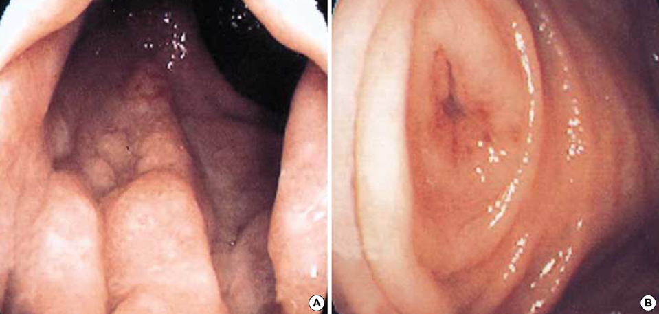
Fig. 1 Colonoscopic findings at admission. (A) Colonoscopy showing granular & reddish mucosal thickening in the rectum just above anus. (B) Colonoscopy showing reddish inflammatory mucosal swelling in the appendiceal orifice.
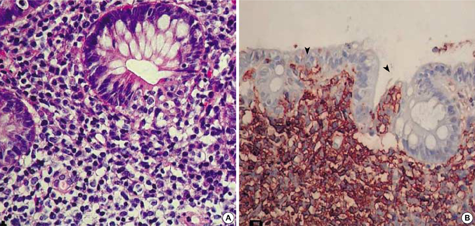
Fig. 2 Histologic findings of biopsy specimens. (A) Neoplastic lymphoid cells infiltrated colonic gland resembling a lymphoepithelial lesion (H&E, ×200). (B) Immunohistochemical stain for CD 20 showing diffuse reaction in the cell membrane. Lymphoepithelial lesions (arrow heads) are also positive for CD20 (×200).
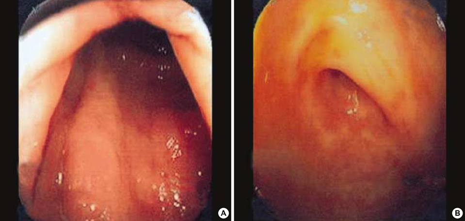
Fig. 3 Colonoscopic findings after treatment. (A) Colonoscopy showing no definitive abnormal lesions in the rectum just above anus. (B) Colonoscopy showing normal appendiceal orifice.
Cited by 3 articles
-
Rare Cause of a Colonic Laterally Spreading Tumor
Sung Min Lee, Dong Hae Chung, Kwang An Kwon
Clin Endosc. 2020;53(4):499-501. doi: 10.5946/ce.2020.197.A Case of Mucosa-Associated Lymphoid Tissue Lymphoma of the Sigmoid Colon Presenting as a Semipedunculated Polyp
Myung Hwan Kim, Jin Tae Jung, Eui Jung Kim, Tae Won Kim, Seon Young Kim, Joong Goo Kwon, Eun Young Kim, Woo Jung Sung
Clin Endosc. 2014;47(2):192-196. doi: 10.5946/ce.2014.47.2.192.MALT (Mucosa-Associated Lymphoid Tissue) Lymphoma of the Colon
Jong Hyeok Park, Jung Hwan Lee
Korean J Gastroenterol. 2010;55(4):213-216. doi: 10.4166/kjg.2010.55.4.213.
Reference
-
1. Isaacson PG, Wright DH. Malignant lymphoma of mucosa-associated lymphoid tissue: A distinctive type of B-cell lymphoma. Cancer. 1983. 52:1410–1416.
Article2. The Non-Hodgkin's Lymphoma Classification Project. A clinical evaluation of the International Lymphoma Study Group classification of non-Hodgkin's lymphoma. Blood. 1997. 89:3909–3918.3. Harris NL, Jaffe ES, Stein H, Banks PM, Chan JK, Cleary ML, Delsol G, De Wolf-Peeters C, Falini B, Gatter KC, Grogan TM, Isaacson PG, Knowles DM, Mason DY, Muller-Hermelink HK, Pileri SA, Piris MA, Ralfkiaer E, Warnke RA. A revised European-American classification of lymphoid neoplasms: a proposal from the International Lymphoma Study Group. Blood. 1994. 84:1361–1392.4. Harris NL, Jaffe ES, Diebold J, Flandrin G, Muller-Hermelink HK, Vardiman J, Lister TA, Bloomfield CD. The World Health Organization classification of neoplastic diseases of the hematopoietic and lymphoid tissues: report of the clinical advisory committee meeting, Airlie House, Virginia, November, 1997. Ann Oncol. 1999. 10:1419–1432.5. Thieblemont C, Berger F, Dumontet C, Moullet I, Bouafia F, Felman P, Salles G, Coiffier B. Mucosa-associated lymphoid tissue lymphoma is a disseminated disease in one third of 158 patients analyzed. Blood. 2000. 95:802–806.
Article6. Radaszkiewicz T, Dragosics B, Bauer P. Gastrointestinal malignant lymphomas of the mucosa-associated lymphoid tissue: Factors relevant to prognosis. Gastroenterology. 1992. 102:1628–1638.
Article7. d'Amore F, Brincker H, Gronbaek K, Thorling K, Pedersen M, Jensen MK, Andersen E, Pedersen NT, Mortensen LS. Non-Hodgkin's lymphoma of the gastrointestinal tract: a population-based analysis of incidence, geographic distribution, clinicopathologic presentation features and prognosis. Danish Lymphoma study group. J Clin Oncol. 1994. 12:1673–1684.8. Hahn JS, Kim YS, Lee YC, Yang WI, Lee SY, Suh CO. Eleven-year experience of low grade lymphoma in Korea (based on REAL classification). Yonsei Med J. 2003. 44:757–770.
Article9. Shepherd NA, Hall PA, Coates PJ, Levinson DA. Primary malignant lymphoma of the colon and rectum. A histopathological and immunohistochemical analysis of 45 cases with clinicopathological correlations. Histopathology. 1988. 12:235–252.
Article10. Schmid C, Vazquez JJ, Diss TC, Isaacson PG. Primary B-cell mucosa associated lymphoid tissue lymphoma presenting as a solitary colorectal polyp. Histopathology. 1994. 24:357–362.11. Park JS, Kim WH, Baek HJ, Kim SH, Kim BS, Woo SG, Park JH, Kim IS. A case of mucosa-associated lymphoid tissue (MALT) lymphoma of the large intestine diagnosed by sigmoidoscopy. Korean J Gastrointest Endosc. 2001. 233:122–126.12. Zucca E, Bertoni F, Roggero E, Cavalli F. The gastric marginal zone B-cell lymphoma of MALT type. Blood. 2000. 96:410–419.
Article13. Zucca E, Roggero E, Pileri S. B-cell lymphoma of MALT type: a review with special emphasis on diagnostic and management problems of low-grade gastric tumours. Br J Haematol. 1998. 100:3–14.
Article14. Zucca E, Cavalli F. Are antibiotics the treatment of choice for gastric lymphoma? Curr Hematol Rep. 2004. 3:11–16.15. Takada M, Ichihara T, Fukumoto S, Nomura H, Kuroda Y. Laparoscopy-assisted colon resection for mucosa-associated lymphoid tissue (MALT) lymphoma in the cecum. Hepatogastroenterology. 2003. 50:1003–1005.16. Rhee JC, Lee HY, Rhee PL, Kim JJ, Paik SW, Koh KC, Go JH. Endoscopic findings of gastric mucosa-associated lymphoid tissue (MALT) lymphoma. Korean J Gastrointest Endosc. 1997. 17:125–131.
- Full Text Links
-
- Actions
-
Cited
- CITED
-
- Close
- Share
-
- Similar articles
-
- A Case of Mucosa-Associated Lymphoid Tissue Lymphoma of Colon as Multiple Large Polypoid Lesions
- Successful Endoscopic Resection of Residual Colonic Mucosa-Associated Lymphoid Tissue Lymphoma after Polypectomy
- A Case of Primary Rectal Colon Mucosa associated Lymphoid Tissue Lymphoma
- A case report of the Pulmonary Malignant Lymphomaof the mucosa-associated lymphoid tissue(MALT)
- A Case of Mucosa-Associated Lymphoid Tissue Lymphoma of the Sigmoid Colon Presenting as a Semipedunculated Polyp
