Hemoperitoneum Caused by Hepatic Necrosis and Rupture Following a Snakebite: a Case Report with Rare CT Findings and Successful Embolization
- Affiliations
-
- 1Department of Radiology, Asan Foundation, GangNeung Asan Hospital, University of Ulsan College of Medicine, Gangneung, Korea. smjung@gnah.co.kr
- 2Department of Thoracic and Cardiovascular Surgery, Asan Foundation, GangNeung Asan Hospital, University of Ulsan College of Medicine, Gangneung, Korea.
- 3Department of General Surgery, Asan Foundation, GangNeung Asan Hospital, University of Ulsan College of Medicine, Gangneung, Korea.
- KMID: 1089446
- DOI: http://doi.org/10.3348/kjr.2007.8.6.556
Abstract
- We report the computed tomographic and angiographic findings in the case of a recently obtained successful clinical outcome after embolization of the hepatic artery in the case of a snakebite causing hemoperitoneum associated with hepatic necrosis and rupture with active bleeding.
MeSH Terms
-
Aged, 80 and over
Contrast Media/administration & dosage
Embolization, Therapeutic/*methods
Female
Fibrin Foam/therapeutic use
Follow-Up Studies
Hemoglobins
Hemoperitoneum/*etiology/therapy
Hemorrhage/etiology/therapy
Hepatic Artery/radiography
Humans
Korea
Liver/*injuries/pathology/radiography
Massive Hepatic Necrosis/complications/*etiology/therapy
Radiographic Image Enhancement/methods
Rupture, Spontaneous
Snake Bites/*complications
Tomography, X-Ray Computed/*methods
Treatment Outcome
Viper Venoms/adverse effects
Figure
-
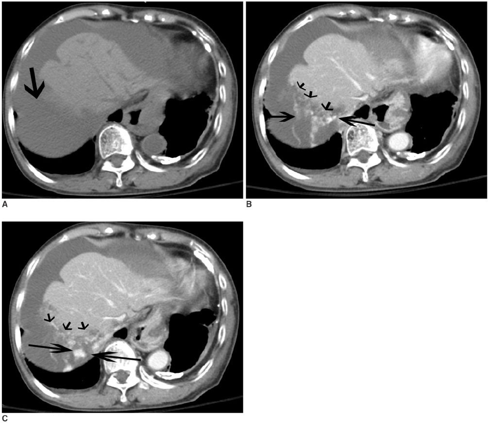
Fig. 1 Unenhanced axial CT scan (A) shows a hematoma in the perihepatic space (arrow). Enhanced CT scans (B, C) show an irregular interface between the hepatic parenchyma and perihepatic hematoma, which presumably represents the site of hepatic rupture (short arrows) and multiple active contrast extravasations (long arrows).
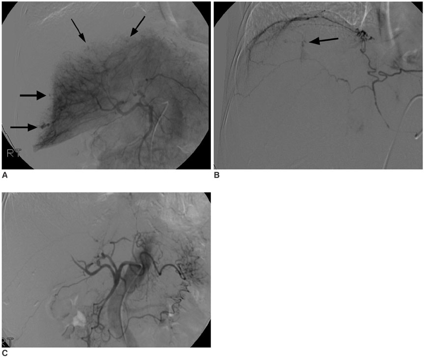
Fig. 2 Hepatic angiogram (A) shows multiple contrast extravasations in the peripheral liver (arrows). A right inferior phrenic angiogram (B) shows the focal contrast extravasation (arrow). A post-embolization angiogram with gelatin sponge (C) shows no evidence of hemorrhage.
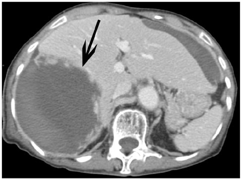
Fig. 3 A follow-up enhanced CT scan after embolization two weeks later shows a large post-hemorrhagic pseudocyst formation (arrow).
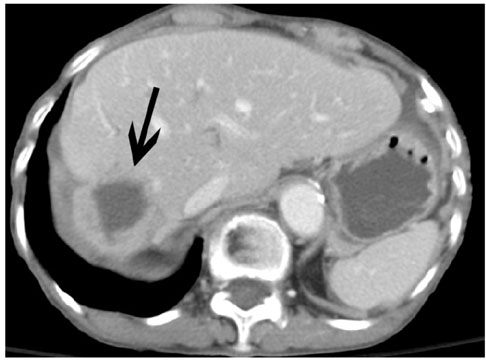
Fig. 4 A follow-up enhanced CT scan after embolization performed 16 weeks later shows a decrease in size of the post-hemorrhagic pseudocyst as well as gradual atrophy of the right lobe of the liver (arrow).
Reference
-
1. Chippaux JP. Snake-bites: appraisal of the global situation. Bull World Health Organ. 1988. 76:515–524.2. White J. Dart R, editor. Overview of venomous snakes of the world. Medical Toxicology. 2004a. Lippincott: Williams and Wilkins;1543–1559.3. Rathod K, Sheth R, Chavhan G, Asrani A, Raut A. Hemoperitoneum complicating snake bite: rare CT features. Abdom Imaging. 2003. 28:820–821.4. Iwakiri R, Fujimoto K, Hirano M, Hisatsugu T, Nojiri I, Sakemi T. Snake-strike-induced ischemic colitis with colonic stricture complicated by disseminated intravascular coagulation. South Med J. 1995. 88:1084–1085.5. Rosenthal R, Meier J, Koelz A, Müller C, Wegmann W, Vogelbach P. Intestinal ischemia after buchmaster (Lachesis muta) snakebite- a case report. Toxicon. 2002. 40:217–220.6. Lee BC, Hwang SH, Bae JC, Kwon SB. Brainstem infarction following Korean viper bite. Neurology. 2001. 56:1244–1245.7. Nunes JO, Turner MA, Fulcher AS. Abdominal imaging feature of HELLP syndrome: A 10-year retrospective review. AJR Am J Roentgenol. 2005. 185:1205–1210.8. Lee JW, Seu JH, Rhee IK, Jin I, Kawamura Y, Park W. Purification and characterization of brevinase, a heterogeneous two-chain fibrinolytic enzyme from the venom of Korean snake, Agkistrodon blomhoffii brevicaudus. Biochem Biophys Res Commun. 1999. 260:665–670.9. Li ZY, Wu XW, Yu TF, Lian EC. Purification and characterization of lupus anticoagulant like protein form Agkistrodon halys brevicaudus venom. Thromb Haemost. 1996. 76:791–797.10. Xu X, Wang Y, Wei C, Zhu X. Study on the action mechanism of hemorrhagin I from Agkidtrodon acutus venom. Adv Exp Med Biol. 1996. 391:361–366.
- Full Text Links
-
- Actions
-
Cited
- CITED
-
- Close
- Share
-
- Similar articles
-
- Hepatic Hemangioma Rupture Caused by Blunt Trauma
- Spontaneous hepatic haemangioma rupture and hemoperitoneum: a double problem with a single stage interventional radiology solution
- Gelatin Sponge Particle Embolization of Spontaneously Ruptured Intrahepatic Arterial Aneurysms in a Patient with Polyarteritis Nodosa: A Case Report
- Embolotherapy of Ruptured Gastroepiploico-Colic Communicating Artery with Median Arcuate Ligament Syndrome: A Case Report
- Hepatic Rupture Caused by Hemolysis, Elevated Liver Enzyme, and Low Platelet Count Syndrome: A Case Report with Computed Tomographic and Conventional Angiographic Findings
