Comparative immunohistochemical characterization of canine seminomas and Sertoli cell tumors
- Affiliations
-
- 1Department of Veterinary Pathobiology, Small Animal Tumor Diagnostic Center, College of Veterinary Medicine, Konkuk University, Seoul 143-701, Korea. jsur@konkuk.ac.kr
- 2Department of Animal Science, Korea National Agricultural College, Hwaseong 445-760, Korea.
- KMID: 1089339
- DOI: http://doi.org/10.4142/jvs.2009.10.1.1
Abstract
- Primary testicular tumors are the most common causes of cancer in male dogs. Overall, the majority of canine patients should be cured by testicular surgery. However, tumor markers are not well-known in veterinary medicine. We sought to determine using immunohistochemistry whether the combined human testicular tumor markers (placental alkaline phosphatase, OCT3/4, CD30, alpha-fetoprotein, inhibin-alpha, vimentin, c-KIT, and desmin) are expressed in canine seminomas and Sertoli cell tumors (SCTs). We examined 35 canine testicular tumors, 20 seminomas and 15 SCTs. c-KIT was expressed markedly in canine seminomas. Both inhibin-alpha and vimentin were expressed significantly in canine SCTs. The results of this study demonstrate differences and similarities between tumor marker expression of testicular tumors in dogs and humans. All the main markers in current routine use are discussed as well as potential useful markers for benign and malignant tumors, and tumor progression.
Keyword
MeSH Terms
Figure
-

Fig. 1 Immunohistochemical markers in canine seminoma. (A) Seminomas consisted of aggregates of germ cells that filled the affected seminiferous tubules. The tumor cells were large and polyhedral with vesicular nuclei and prominent nucleoli. H&E stain. (B-F) Positive signals to tumor cells; (B) alpha-fetoprotein, (C) inhibin-alpha, (D) vimentin, (E) desmin and (F) c-KIT. Immunostain and counterstain with Harris hematoxylin. Scale bars = A: 350 µm, B-F: 140 µm.
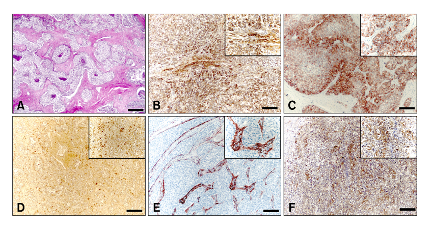
Fig. 2 Immunohistochemical markers in canine Sertoli cell tumors. (A) Cells within the tumor resemble Sertoli cells that normally populate the seminiferous tubules and are arranged into sheets or tubules separated by fibrous connective tissues. H&E stain. (B-F) Positive signals to tumor cells; (B) alpha-fetoprotein, (C) inhibin-alpha, (D) vimentin, (E) desmin and (F) c-KIT. Immunostain and counterstain with Harris hematoxylin. Scale bars = A: 350 µm, B-F: 140 µm.
Reference
-
1. Biermann K, Klingmüller D, Koch A, Pietsch T, Schorle H, Büttner R, Zhou H. Diagnostic value of markers M2A, OCT3/4, AP-2gamma, PLAP and c-KIT in the detection of extragonadal seminomas. Histopathology. 2006. 49:290–297.
Article2. Cheng L. Establishing a germ cell origin for metastatic tumors using OCT4 immunohistochemistry. Cancer. 2004. 101:2006–2010.
Article3. Cheng L, Thomas A, Roth LM, Zheng W, Michael H, Karim FW. OCT4: a novel biomarker for dysgerminoma of the ovary. Am J Surg Pathol. 2004. 28:1341–1346.4. Debora J, Junqi Q, David GB. Dabbs DJ, editor. Immunohistology of the prostate, bladder, testis and kidney. Diagnostic Immunohistochemistry. 2002. 2nd ed. New York: Churchill Livingstone;75–86.5. De Jong FH, Grootenhuis AJ, Steenbergen J, van Sluijs FJ, Foekens JA, ten Kate FJ, Oosterhuis JW, Lamberts SW, Klijn JG. Inhibin immunoreactivity in gonadal and non-gonadal tumors. J Steroid Biochem Mol Biol. 1990. 37:863–866.
Article6. De Vico G, Papparella S, Di Guardo G. Number and size of silver-stained nucleoli (Ag-NOR clusters) in canine seminomas: correlation with histological features and tumour behaviour. J Comp Pathol. 1994. 110:267–273.
Article7. Goto T, Adjaye J, Rodeck CH, Monk M. Identification of genes expressed in human primordial germ cells at the time of entry of the female germ line into meiosis. Mol Hum Reprod. 1999. 5:851–860.
Article8. Grieco V, Riccardi E, Rondena M, Ciampi V, Finazzi M. Classical and spermatocytic seminoma in the dog: histochemical and immunohistochemical findings. J Comp Pathol. 2007. 137:41–46.
Article9. Grootenhuis AJ, van Sluijs FJ, Klaij IA, Steenbergen J, Timmerman MA, Bevers MM, Dieleman SJ, de Jong FH. Inhibin, gonadotrophins and sex steroids in dogs with Sertoli cell tumours. J Endocrinol. 1990. 127:235–242.
Article10. Hansis C, Grifo JA, Krey LC. Oct-4 expression in inner cell mass and trophectoderm of human blastocysts. Mol Hum Reprod. 2000. 6:999–1004.
Article11. Iczkowski KA, Butler SL. New immunohistochemical markers in testicular tumors. Anal Quant Cytol Histol. 2006. 28:181–187.12. Izquierdo MA, Van der Valk P, Van Ark-Otte J, Rubio G, Germa-Lluch JR, Ueda R, Scheper RJ, Takahashi T, Giaccone G. Differential expression of the c-kit proto-oncogene in germ cell tumours. J Pathol. 1995. 177:253–258.
Article13. Jacobsen GK, Jacobsen M. Alpha-fetoprotein (AFP) and human chorionic gonadotropin (HCG) in testicular germ cell tumours. A prospective immunohistochemical study. Acta Pathol Microbiol Immunol Scand [A]. 1983. 91:165–176.14. Jones TD, Ulbright TM, Eble JN, Baldridge LA, Cheng L. OCT4 staining in testicular tumors: a sensitive and specific marker for seminoma and embryonal carcinoma. Am J Surg Pathol. 2004. 28:935–940.15. Kawakami E, Hirano T, Hori T, Tsutsui T. Testicular superoxide dismutase activity, heat shock protein 70 concentration and blood plasma inhibin-alpha concentration of dogs with a Sertoli cell tumor in a unilateral cryptorchid testis. J Vet Med Sci. 2007. 69:1259–1262.
Article16. Kommoss F, Oliva E, Bittinger F, Kirkpatrick CJ, Amin MB, Bhan AK, Young RH, Scully RE. Inhibin-alpha CD99, HEA125, PLAP, and chromogranin immunoreactivity in testicular neoplasms and the androgen insensitivity syndrome. Hum Pathol. 2000. 31:1055–1061.
Article17. Koshida K, Uchibayashi T, Yamamoto H, Hirano K. Significance of placental alkaline phosphatase (PLAP) in the monitoring of patients with seminoma. Br J Urol. 1996. 77:138–142.
Article18. Lau SK, Weiss LM, Chu PG. D2-40 immunohistochemistry in the differential diagnosis of seminoma and embryonal carcinoma: a comparative immunohistochemical study with KIT (CD117) and CD30. Mod Pathol. 2007. 20:320–325.
Article19. Leroy X, Augusto D, Leteurtre E, Gosselin B. CD30 and CD117 (c-kit) used in combination are useful for distinguishing embryonal carcinoma from seminoma. J Histochem Cytochem. 2002. 50:283–285.
Article20. McCluggage WG, Shanks JH, Whiteside C, Maxwell P, Banerjee SS, Biggart JD. Immunohistochemical study of testicular sex cord-stromal tumors, including staining with anti-inhibin antibody. Am J Surg Pathol. 1998. 22:615–619.
Article21. McEntee K. McEntee K, editor. Scrotum, Spermatic cord and testis proliferative lesions. Reproductive Pathology of Domestic Mammals. 1990. San Diego: Academic Press;279–300.
Article22. McLachlan NJ, Kennedy PC. Meuten DJ, editor. Tumors of the genital system. Tumors in Domestic Animals. 2002. 4th ed. Ames: Iowa State University Press;561–573.23. Miller MA, Hartnett SE, Ramos-Vara JA. Interstitial cell tumor and Sertoli cell tumor in the testis of a cat. Vet Pathol. 2007. 44:394–397.
Article24. Mooney EE, Nogales FF, Bergeron C, Tavassoli FA. Retiform Sertoli-Leydig cell tumours: clinical, morphological and immunohistochemical findings. Histopathology. 2002. 41:110–117.
Article25. Mostofi FK, Sesterhenn IA. Pathology of germ cell tumors of testes. Prog Clin Biol Res. 1985. 203:1–34.26. Nakai Y, Nonomura N, Oka D, Shiba M, Arai Y, Nakayama M, Inoue H, Nishimura K, Aozasa K, Mizutani Y, Miki T, Okuyama A. KIT (c-KIT oncogene product) pathway is constitutively activated in human testicular germ cell tumors. Biochem Biophys Res Commun. 2005. 337:289–296.
Article27. Nikolaou M, Valavanis C, Aravantinos G, Fountzilas G, Tamvakis N, Lekka I, Arapantoni-Dadioti P, Zizi A, Ghiconti I, Economopoulos T, Pectasides D. KIT expression in male germ cell tumors. Anticancer Res. 2007. 27:1685–1688.28. Owston MA, Ramos-Vara JA. Histologic and immunohistochemical characterization of a testicular mixed germ cell sex cord-stromal tumor and a leydig cell tumor in a dog. Vet Pathol. 2007. 44:936–943.
Article29. Pera MF, Herszfeld D. Differentiation of human pluripotent teratocarcinoma stem cells induced by bone morphogenetic protein-2. Reprod Fertil Dev. 1998. 10:551–555.
Article30. Peters MA, Teerds KJ, van der Gaag I, de Rooij DG, van Sluijs FJ. Use of antibodies against LH receptor, 3beta-h droxysteroid dehydrogenase and vimentin to characterize different types of testicular tumour in dogs. Reproduction. 2001. 121:287–296.31. Rajpert-De Meyts E. Recent advances and future directions in research on testicular germ cell cancer. Int J Androl. 2007. 30:192–197.
Article32. Rosai J. Rosai and Ackerman's Surgical Pathology. 2004. 4th ed. Edinburgh: Mosby;1412–1456.33. Sung MT, Jones TD, Beck SD, Foster RS, Cheng L. OCT4 is superior to CD30 in the diagnosis of metastatic embryonal carcinomas after chemotherapy. Hum Pathol. 2006. 37:662–667.
Article34. Taniyama H, Hirayama K, Nakada K, Numagami K, Yaosaka N, Kagawa Y, Izumisawa Y, Nakade T, Tanaka Y, Watanabe G, Taya K. Immunohistochemical detection of inhibin-alpha, -betaB, and -betaA chains and 3beta-ydroxysteroid dehydrogenase in canine testicular tumors and normal testes. Vet Pathol. 2001. 38:661–666.
Article35. Teng LH, Lu DH, Xu QZ, Fu YJ, Yang H, He ZL. Expression and diagnostic significance of OCT4, CD117 and CD30 in germ cell tumors. Zhonghua Bing Li Xue Za Zhi. 2005. 34:711–715.36. Ulbright TM, Amin MB, Young RH. Tumors of the Testis, Adnexa, Spermatic Cord, and Scrotum. 1999. 3rd ed. Washington DC: Armed Forces Institute of Pathology;59–85.
- Full Text Links
-
- Actions
-
Cited
- CITED
-
- Close
- Share
-
- Similar articles
-
- Testis tumor: A clinical experience of 20 years
- Ovarian serous cystadenoma associated with Sertoli-Leydig cell tumor: a case report
- A Case of Sertoli-Leydig Cell Tumor
- A Case of Giant Sertoli-Leydig Cell Tumor of the Ovary with Masculinization
- A Case of Sertoli-Leydig Cell Tumor Combined with Dermoid Cyst
