Korean J Radiol.
2009 Apr;10(2):101-105. 10.3348/kjr.2009.10.2.101.
Ultrasonographic Findings of Medullary Thyroid Carcinoma: a Comparison with Papillary Thyroid Carcinoma
- Affiliations
-
- 1Department of Radiology, College of Medicine, The Catholic University of Korea, Korea. bumrad@catholic.ac.kr
- 2Department of Internal Medicine, College of Medicine, The Catholic University of Korea, Korea.
- 3Department of Pathology, College of Medicine, The Catholic University of Korea, Korea.
- KMID: 1088719
- DOI: http://doi.org/10.3348/kjr.2009.10.2.101
Abstract
OBJECTIVE
This study was designed to evaluate the ultrasonographic (US) findings of medullary thyroid carcinoma (MTC) as compared to findings for papillary thyroid carcinoma (PTC).
MATERIALS AND METHODS
The study included 21 cases of MTC that were surgically diagnosed between 2002 and 2007 and 114 cases of PTC that were diagnosed in 2007. Two radiologists reached a consensus in the evaluation of the US findings. The US findings were classified as recommended by the Thyroid Study Group of the Korean Society of Neuroradiology and Head and Neck Radiology (KSNHNR) and each nodule was identified as suspicious malignant, indeterminate or probably benign. The findings of medullary and papillary carcinomas were compared with use of the chi-squared test.
RESULTS
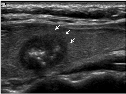
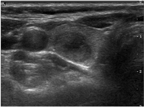
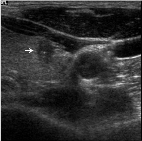
The common US findings for MTCs were solid internal content (91%), an ovoid to round shape (57%), marked hypoechogenicity (52%) and calcifications (52%). Among the 21 cases of MTC nodules, 17 (81%) were classified as suspicious malignant nodules. The mean size (longest diameter) of MTC nodules was 19 +/- 13.9 mm and the mean size (longest diameter) of PTC nodules was 11 +/- 7.4 mm; this difference was statistically significant (p < 0.05). An ovoid to round shape was more prevalent for MTC lesions than for PTC lesions (p < 0.05).
CONCLUSION
The US criteria for suspicious malignant nodules as recommended by the Thyroid Study Group of the KSNHNR correspond to most MTC cases. The US findings for MTC are not greatly different from PTC except for the prevalence of an ovoid to round shape.
Keyword
MeSH Terms
Figure
Cited by 1 articles
-
Cystic Medullary Thyroid Carcinoma: A Case of Undergoing Endoscopic Thyroid Lobectomy
Dong Hae Chung, Jae Yeon Seok, Yoo Seung Chung, Eun Mee Oh, Jung Won Ryu, Young Don Lee
Korean J Endocr Surg. 2015;15(1):15-19. doi: 10.16956/kjes.2015.15.1.15.
Reference
-
1. McCook TA, Putman CE, Dale JK, Wells SA. Medullary carcinoma of the thyroid: radiographic features of a unique tumor. AJR Am J Roentgenol. 1982. 139:149–155.2. Melvin KE, Tashjian AH Jr. The syndrome of excessive thyrocalcitonin produced by medullary carcinoma of the thyroid. Proc Natl Acad Sci USA. 1968. 59:1216–1222.3. Busnardo B, Girelli ME, Simioni N, Nacamulli D, Busetto E. Nonparallel patterns of calcitonin and carcinoembryonic antigen levels in the follow-up of medullary thyroid carcinoma. Cancer. 1984. 53:278–285.4. Papi G, Corsello SM, Cioni K, Pizzini AM, Corrado S, Carapezzi C, et al. Value of routine measurement of serum calcitonin concentrations in patients with nodular thyroid disease: a multicenter study. J Endocrinol Invest. 2006. 29:427–437.5. You YN, Lakhani V, Wells SA Jr, Moley JF. Medullary thyroid cancer. Surg Oncol Clin N Am. 2006. 15:639–660.6. Moon WJ, Jung SL, Lee JH, Na DG, Baek JH, Lee YH, et al. Benign and malignant thyroid nodules: US differentiation-multicenter retrospective study. Radiology. 2008. 247:762–777.7. Frates MC, Benson CB, Charboneau JW, Cibas ES, Clark OH, Coleman BG, et al. Management of thyroid nodules detected at US: Society of Radiologists in Ultrasound consensus conference statement. Radiology. 2005. 237:794–800.8. Iannuccilli JD, Cronan JJ, Monchik JM. Risk for malignancy of thyroid nodules as assessed by sonographic criteria: the need for biopsy. J Ultrasound Med. 2004. 23:1455–1464.9. Kim EK, Park CS, Chung WY, Oh KK, Kim DI, Lee JT, et al. New sonographic criteria for recommending fine-needle aspiration biopsy of nonpalpable solid nodules of the thyroid. AJR Am J Roentgenol. 2002. 178:687–691.10. Shimura H, Haraguchi K, Hiejima Y, Fukunari N, Fujimoto Y, Katagiri M, et al. Distinct diagnostic criteria for ultrasonographic examination of papillary thyroid carcinoma: a multicenter study. Thyroid. 2005. 15:251–258.11. Chan BK, Desser TS, McDougall IR, Weigel RJ, Jeffrey RB Jr. Common and uncommon sonographic features of papillary thyroid carcinoma. J Ultrasound Med. 2003. 22:1083–1090.12. Pellitteri PK, McCaffrey TV. Tumor and tumor-like lesion in thyroid endocrine surgery of the head and neck. 2002. 1st ed. Pennsylvania: Singular publish group;21–47.13. Jeh SK, Jung SL, Kim BS, Lee YS. Evaluating the degree of conformity of papillary carcinoma and follicular carcinoma to the reported ultrasonographic findings of malignant thyroid tumor. Korean J Radiol. 2007. 8:192–197.14. Gorman B, Charboneau JW, James EM, Reading CC, Wold LE, Grant CS, et al. Medullary thyroid carcinoma: role of highresolution US. Radiology. 1987. 162:147–150.15. Saller B, Moeller L, Görges R, Janssen OE, Mann K. Role of conventional ultrasound and color Doppler sonography in the diagnosis of medullary thyroid carcinoma. Exp Clin Endocrinol Diabetes. 2002. 110:403–407.16. Williams ED. Histogenesis of medullary carcinoma of the thyroid. J Clin Pathol. 1966. 19:114–118.17. Hoang JK, Lee WK, Lee M, Johnson D, Farrell S. US features of thyroid malignancy: pearls and pitfalls. Radiographics. 2007. 27:847–860.
- Full Text Links
- Actions
-
Cited
- CITED
-
- Close
- Share
- Similar articles
-
- Concurrent Medullay and Papillary Carcinoma of the Thyroid
- Concurrent Papillary and Medullary Carcinoma of the Thyroid Gland
- A Case of Concurrent Papillary and Medullary Thyroid Carcinomas Detected as Recurrent Medullary Carcinoma after Initial Surgery for Papillary Carcinoma
- Medullary and Papillary Thyroid Carcinoma as a Collision Tumor: Report of Five Cases
- Ultrasonographic Findings of Papillary Thyroid Cancer with or without Hashimoto's Thyroiditis


