Usefulness of the CAD System for Detecting Pulmonary Nodule in Real Clinical Practice
- Affiliations
-
- 1Department of Radiology and Center for Imaging Science, Samsung Medical Center, Sungkyunkwan University School of Medicine, Seoul 135-710, Korea. mj1.chung@samsung.com
- 2Department of Surgery, Samsung Medical Center, Sungkyunkwan University School of Medicine, Seoul 135-710, Korea.
- 3Department of Radiology, Jeju National University College of Medicine, Jeju 690-716, Korea.
- KMID: 1088560
- DOI: http://doi.org/10.3348/kjr.2011.12.2.163
Abstract
OBJECTIVE
We wanted to evaluate the usefulness of the computer-aided detection (CAD) system for detecting pulmonary nodules in real clinical practice by using the CT images.
MATERIALS AND METHODS
Our Institutional Review Board approved our retrospective study with a waiver of informed consent. This study included 166 CT examinations that were performed for the evaluation of pulmonary metastasis in 166 patients with colorectal cancer. All the CT examinations were interpreted by radiologists and they were also evaluated by the CAD system. All the nodules detected by the CAD system were evaluated with regard to whether or not they were true nodules, and they were classified into micronodules (MN, diameter < 4 mm) and significant nodules (SN, 4 < or = diameter < or = 10 mm). The radiologic reports and CAD results were compared.
RESULTS
The CAD system helped detect 426 nodules; 115 (27%) of the 426 nodules were classified as true nodules and 35 (30%) of the 115 nodules were SNs, and 83 (72%) of the 115 were not mentioned in the radiologists' reports and three (4%) of the 83 nodules were non-calcified SNs. One of three non-calcified SNs was confirmed as a metastatic nodule. According to the radiologists' reports, 60 true nodules were detected, and 28 of the 60 were not detected by the CAD system.
CONCLUSION
Although the CAD system missed many SNs that are detected by radiologists, it helps detect additional nodules that are missed by the radiologists in real clinical practice. Therefore, the CAD system can be useful to support a radiologist's detection performance.
MeSH Terms
Figure
-

Fig. 1 57-year-old man who underwent transanal endoscopic microsurgery for rectal cancer four years before chest CT scan. Candidate lesion is marked (arrow) on captured image from computer-aided detection. This was actually branch of right superior pulmonary vein.

Fig. 2 55-year-old man who underwent low anterior resection for rectal cancer six months before chest CT scan. A. On axial CT image, small non-calcified lung nodule (diameter = 5.4 mm) is noted in right minor fissure (arrow). B. On captured image from computer-aided detection, nodule is marked as C1 candidate lesion (arrow). This nodule was detected by both computer-aided detection system and radiologist. C. On axial CT image performed one year later, nodule shows no interval change of size and shape (arrow). Note newly appeared malignant pleural effusion in left hemithorax.
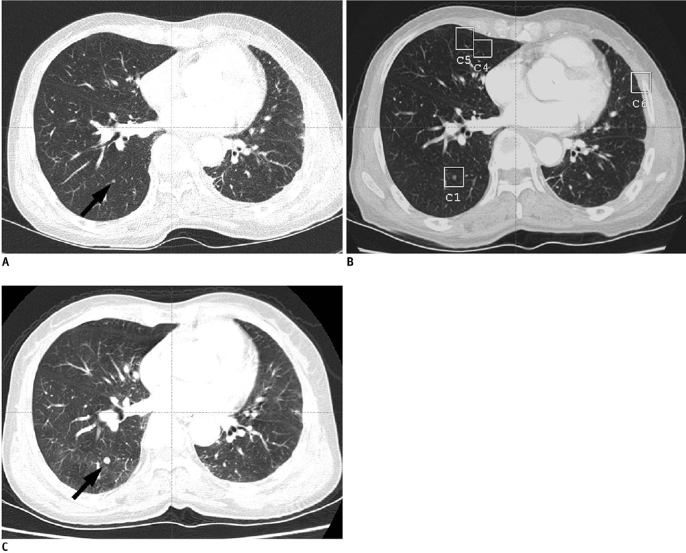
Fig. 3 61-year-old woman who underwent right hemicolectomy for colon cancer one year before chest CT scan. A. On axial CT image, small lung nodule (diameter = 4.0 mm) is noted in right lower lobe (arrow). This non-calcified significant nodule was missed in official report. B. On captured image from computer-aided detection, same nodule is marked as C1, candidate lesion. C. On axial CT image performed four months later, nodule demonstrated interval increase in size (arrow). Metastatic adenocarcinoma was confirmed by video-assisted thoracoscopic surgery metastasectomy.
Cited by 1 articles
-
An Engineering View on Megatrends in Radiology: Digitization to Quantitative Tools of Medicine
Namkug Kim, Jaesoon Choi, Jaeyoun Yi, Seungwook Choi, Seyoun Park, Yongjun Chang, Joon Beom Seo
Korean J Radiol. 2013;14(2):139-153. doi: 10.3348/kjr.2013.14.2.139.
Reference
-
1. Ambiru S, Miyazaki M, Ito H, Nakagawa K, Shimizu H, Kato A, et al. Resection of hepatic and pulmonary metastases in patients with colorectal carcinoma. Cancer. 1998. 82:274–278.2. Girard P, Ducreux M, Baldeyrou P, Rougier P, Le Chevalier T, Bougaran J, et al. Surgery for lung metastases from colorectal cancer: analysis of prognostic factors. J Clin Oncol. 1996. 14:2047–2053.3. McAfee MK, Allen MS, Trastek VF, Ilstrup DM, Deschamps C, Pairolero PC. Colorectal lung metastases: results of surgical excision. Ann Thorac Surg. 1992. 53:780–785. discussion 785-786.4. Yano T, Hara N, Ichinose Y, Yokoyama H, Miura T, Ohta M. Results of pulmonary resection of metastatic colorectal cancer and its application. J Thorac Cardiovasc Surg. 1993. 106:875–879.5. Armato SG 3rd, Li F, Giger ML, MacMahon H, Sone S, Doi K. Lung cancer: performance of automated lung nodule detection applied to cancers missed in a CT screening program. Radiology. 2002. 225:685–692.6. Awai K, Murao K, Ozawa A, Komi M, Hayakawa H, Hori S, et al. Pulmonary nodules at chest CT: effect of computer-aided diagnosis on radiologists' detection performance. Radiology. 2004. 230:347–352.7. Doi K. Current status and future potential of computer-aided diagnosis in medical imaging. Br J Radiol. 2005. 78:S3–S19.8. Ko JP, Naidich DP. Computer-aided diagnosis and the evaluation of lung disease. J Thorac Imaging. 2004. 19:136–155.9. Rubin GD, Lyo JK, Paik DS, Sherbondy AJ, Chow LC, Leung AN, et al. Pulmonary nodules on multi-detector row CT scans: performance comparison of radiologists and computer-aided detection. Radiology. 2005. 234:274–283.10. Yuan R, Vos PM, Cooperberg PL. Computer-aided detection in screening CT for pulmonary nodules. AJR Am J Roentgenol. 2006. 186:1280–1287.11. Choi EJ, Jin GY, Han YM, Lee YS, Kweon KS. Solitary pulmonary nodule on helical dynamic CT scans: analysis of the enhancement patterns using a computer-aided diagnosis (CAD) system. Korean J Radiol. 2008. 9:401–408.12. Lee JY, Chung MJ, Yi CA, Lee KS. Ultra-low-dose MDCT of the chest: influence on automated lung nodule detection. Korean J Radiol. 2008. 9:95–101.13. Ozekes S, Osman O, Ucan ON. Nodule detection in a lung region that's segmented with using genetic cellular neural networks and 3D template matching with fuzzy rule based thresholding. Korean J Radiol. 2008. 9:1–9.14. Armato SG 3rd, Giger ML, MacMahon H. Automated detection of lung nodules in CT scans: preliminary results. Med Phys. 2001. 28:1552–1561.15. Giger ML, Bae KT, MacMahon H. Computerized detection of pulmonary nodules in computed tomography images. Invest Radiol. 1994. 29:459–465.16. Gurcan MN, Sahiner B, Petrick N, Chan HP, Kazerooni EA, Cascade PN, et al. Lung nodule detection on thoracic computed tomography images: preliminary evaluation of a computer-aided diagnosis system. Med Phys. 2002. 29:2552–2558.17. Ko JP, Betke M. Chest CT: automated nodule detection and assessment of change over time--preliminary experience. Radiology. 2001. 218:267–273.18. Wormanns D, Fiebich M, Saidi M, Diederich S, Heindel W. Automatic detection of pulmonary nodules at spiral CT: clinical application of a computer-aided diagnosis system. Eur Radiol. 2002. 12:1052–1057.19. Lee IJ, Gamsu G, Czum J, Wu N, Johnson R, Chakrapani S. Lung nodule detection on chest CT: evaluation of a computer-aided detection (CAD) system. Korean J Radiol. 2005. 6:89–93.20. Davis SD. CT evaluation for pulmonary metastases in patients with extrathoracic malignancy. Radiology. 1991. 180:1–12.
- Full Text Links
-
- Actions
-
Cited
- CITED
-
- Close
- Share
-
- Similar articles
-
- Studies and Real-World Experience Regarding the Clinical Application of Artificial Intelligence Software for Lung Nodule Detection
- A Computer-Aided Diagnosis for Evaluating Lung Nodules on Chest CT: the Current Status and Perspective
- Automated Lung Nodule Detection at Low-Dose CT: Preliminary Experience
- Deep Learning-Based Computer-Aided Diagnosis in Coronary Artery Calcium-Scoring CT for Pulmonary Nodule Detection: A Preliminary Study
- Lung Nodule Detection on Chest CT: Evaluation of a Computer-Aided Detection (CAD) System
