Korean J Ophthalmol.
2008 Dec;22(4):255-258. 10.3341/kjo.2008.22.4.255.
Upper Eyelid Retraction After Periorbital Trauma
- Affiliations
-
- 1Department of Ophthalmology, Hallym University Sacred Heart Hospital, Anyang, Korea. bonamd@paran.com
- KMID: 1084214
- DOI: http://doi.org/10.3341/kjo.2008.22.4.255
Abstract
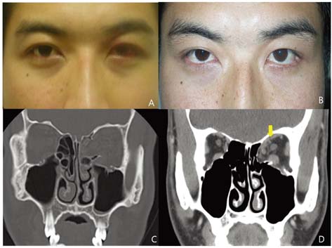
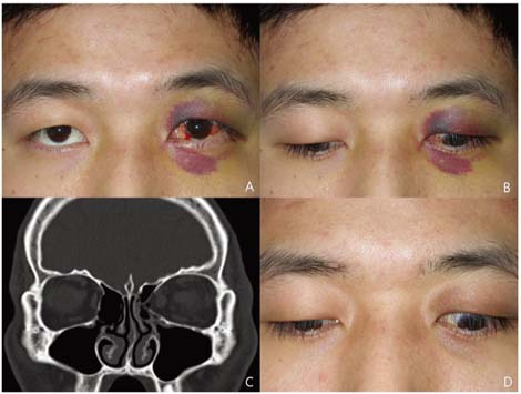
- We report four unusual cases of upper eyelid retraction following periorbital trauma. Four previously healthy patients were evaluated for unilateral upper eyelid retraction following periorbital trauma. A 31-year-old man (Case 1) and a 24-year-old man (Case 2) presented with left upper eyelid retraction which developed after blow-out fractures, a 44-year-old woman (Case 3) presented with left upper eyelid retraction secondary to a periorbital contusion that occurred one week prior, and a 56-year-old man (Case 4) presented with left upper eyelid retraction that developed 1 month after a lower canalicular laceration was sustained during a traffic accident. The authors performed a thyroid function test and orbital computed tomography (CT) in all cases. Thyroid function was normal in all patients, CT showed an adhesion of the superior rectus muscle and superior oblique muscle in the first case and diffuse thickening of the superior rectus muscle and levator complex in the third case. CT showed no specific findings in the second or fourth cases. Upper eyelid retraction due to superior complex adhesion can be considered one of the complications of periorbital trauma.
Keyword
MeSH Terms
Figure
Reference
-
1. Hwang JM, Kim WB. Etiology of eyelid retraction in Koreans. J Korean Ophthalmol Soc. 1998. 39:1069–1076.2. Bartley GB. The differential diagnosis and classification of eyelid retraction. Ophthalmology. 1996. 103:168–176.3. Meyer DR, Wobig JL. Detection of contralateral eyelid retraction associated with blepharoptosis. Ophthalmology. 1992. 99:366–375.4. Putterman AM, Urist MJ. Upper eyelid retraction after blowout fracture. Arch Ophthalmol. 1976. 94:112–116.5. Conway ST. Lid retraction following blow-out fracture of the orbit. Ophthalmic Surg. 1988. 19:279–281.6. Hatt M. Post-traumatic upper eyelid retraction. Klin Monatsbl Augenheilkd. 1985. 186:217–219.7. Mauriello JA Jr, Palydowycz SB. Upper eyelid retraction after retinal detachment repair. Ophthalmic Surg. 1993. 24:694–697.8. Finsterer J. Ptosis: Causes, presentation, and management. Aesthetic Plast Surg. 2003. 27:193–204.9. Bartley GB, Gorman CA. Diagnostic criteria for Graves' ophthalmopathy. Am J Ophthalmol. 1995. 119:792–795.
- Full Text Links
- Actions
-
Cited
- CITED
-
- Close
- Share
- Similar articles
-
- A Case of Congenital Eyelid Retraction
- Surgical Efficacyin the Upper and Lower Eyelid Retraction
- Surgical Treatment of Thyroid-related Upper Eyelid Retraction
- Staged Correction of Cicatricial Upper Eyelid Retraction and Contralateral Upper Eyelid Ptosis
- Acellular Dermal Allograft for the Correction of Eyelid Retraction


