Korean J Ophthalmol.
2011 Dec;25(6):459-462. 10.3341/kjo.2011.25.6.459.
Horner's Syndrome with Abducens Nerve Palsy
- Affiliations
-
- 1Department of Ophthalmology, Ewha Womans University Mokdong Hospital, Seoul, Korea. limkh@ewha.ac.kr
- 2Department of Pathology, Ewha Medical Research Institute, Ewha Womans University School of Medicine, Seoul, Korea.
- KMID: 1031197
- DOI: http://doi.org/10.3341/kjo.2011.25.6.459
Abstract
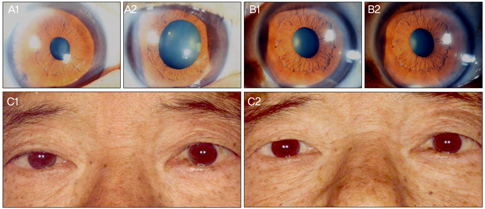
- A 68-year-old male patient presented with a week of sudden diplopia. He had been diagnosed with nasopharyngeal cancer 8 months prior and had undergone chemotherapy with radiotherapy. Eight-prism diopter right esotropia in the primary position and a remarkable limitation in abduction in his right eye were observed. Other pupillary disorders and lid drooping were not found. After three weeks, the marginal reflex distance 1 was 3 mm in the right eye and 5 mm in the left eye. The pupil diameter was 2.5 mm in the right eye, and 3 mm in the left eye under room illumination. Under darkened conditions, the pupil diameter was 3.5 mm in the right eye, and 5 mm in the left eye. After topical application of 0.5% apraclonidine, improvement in the right ptosis and reversal pupillary dilatation were observed. On brain magnetic resonance imaging, enhanced lesions on the right cavernous sinus, both sphenoidal sinuses, and skull base suggested the invasion of nasopharyngeal cancer. Lesions on the cavernous sinus need to be considered in cases of abducens nerve palsy and ipsilateral Horner's syndrome.
MeSH Terms
Figure
Cited by 1 articles
-
Delayed Onset Abducens Nerve Palsy and Horner Syndrome after Treatment of a Traumatic Carotid-cavernous Fistula
Won Jae Kim, Cheol Won Moon, Myung Mi Kim
J Korean Ophthalmol Soc. 2019;60(9):905-908. doi: 10.3341/jkos.2019.60.9.905.
Reference
-
1. Silva MN, Saeki N, Hirai S, Yamaura A. Unusual cranial nerve palsy caused by cavernous sinus aneurysms. Clinical and anatomical considerations reviewed. Surg Neurol. 1999. 52:143–148.2. Rhoton AL Jr. The cavernous sinus, the cavernous venous plexus, and the carotid collar. Neurosurgery. 2002. 51:4 Suppl. S375–S410.3. Ampil FL, Heldmann M, Ibrahim AM, Balfour EL. Involvement of the cavernous sinus by malignant (extracranial) tumour: palliation in six cases without surgery. J Craniomaxillofac Surg. 2000. 28:161–164.4. Tsuda H, Yorinaga Y, Tamada Y, et al. Combination of abducens nerve palsy and ipsilateral postganglionic Horner syndrome as an initial manifestation of uterine cervical cancer. Intern Med. 2009. 48:1457–1460.5. George A, Haydar AA, Adams WM. Imaging of Horner's syndrome. Clin Radiol. 2008. 63:499–505.6. Parkinson D. Bernard, Mitchell, Horner syndrome and others? Surg Neurol. 1979. 11:221–223.7. Kano H, Niranjan A, Kondziolka D, et al. The role of palliative radiosurgery when cancer invades the cavernous sinus. Int J Radiat Oncol Biol Phys. 2009. 73:709–715.8. Curry MP, Newlon JL, Watson DW. Cavernous sinus metastasis from laryngeal squamous cell carcinoma. Otolaryngol Head Neck Surg. 2001. 125:567–568.9. Djalilian HR, Tekin M, Hall WA, Adams GL. Metastatic head and neck squamous cell carcinoma to the brain. Auris Nasus Larynx. 2002. 29:47–54.10. Tsuda H, Ishikawa H, Kishiro M, et al. Abducens nerve palsy and postganglionic Horner syndrome with or without severe headache. Intern Med. 2006. 45:851–855.11. Gutman I, Levartovski S, Goldhammer Y, et al. Sixth nerve palsy and unilateral Horner's syndrome. Ophthalmology. 1986. 93:913–916.12. Tsuda H, Ishikawa H, Asayama K, et al. Abducens nerve palsy and Horner syndrome due to metastatic tumor in the cavernous sinus. Intern Med. 2005. 44:644–646.13. Slamovits TL, Cahill KV, Sibony PA, et al. Orbital fine-needle aspiration biopsy in patients with cavernous sinus syndrome. J Neurosurg. 1983. 59:1037–1042.14. Wilhelm H, Ochsner H, Kopycziok E, et al. Horner's syndrome: a retrospective analysis of 90 cases and recommendations for clinical handling. Ger J Ophthalmol. 1992. 1:96–102.15. Hirao M, Oku H, Sugasawa J, et al. Three cases of abducens nerve palsy accompanied by Horner syndrome. Nihon Ganka Gakkai Zasshi. 2006. 110:520–524.16. Kurihara T. Abducens nerve palsy and ipsilateral incomplete Horner syndrome: a significant sign of locating the lesion in the posterior cavernous sinus. Intern Med. 2006. 45:993–994.17. Ozveren MF, Uchida K, Erol FS, et al. Isolated abducens nerve paresis associated with incomplete Horner's syndrome caused by petrous apex fracture: case report and anatomical study. Neurol Med Chir (Tokyo). 2001. 41:494–498.18. Freedman KA, Brown SM. Topical apraclonidine in the diagnosis of suspected Horner syndrome. J Neuroophthalmol. 2005. 25:83–85.
- Full Text Links
- Actions
-
Cited
- CITED
-
- Close
- Share
- Similar articles
-
- Horner's Syndrome and Contralateral Abducens Nerve Palsy Associated with Zoster Meningitis
- Delayed Onset Abducens Nerve Palsy and Horner Syndrome after Treatment of a Traumatic Carotid-cavernous Fistula
- A Case of Isolated Unilateral Abducens Nerve Palsy Caused by Clival Metastasis from Rectal Cancer
- Abducens Nerve Palsy Associated with Ramsay-Hunt Syndrome
- Fourth Nerve Paresis plus Crossed Horner Syndrome in Acute Leukemia


