J Korean Ophthalmol Soc.
2019 Sep;60(9):905-908. 10.3341/jkos.2019.60.9.905.
Delayed Onset Abducens Nerve Palsy and Horner Syndrome after Treatment of a Traumatic Carotid-cavernous Fistula
- Affiliations
-
- 1Department of Ophthalmology, Yeungnam University College of Medicine, Daegu, Korea. mmk@med.yu.ac.kr
- KMID: 2459138
- DOI: http://doi.org/10.3341/jkos.2019.60.9.905
Abstract
- PURPOSE
We report a patient with delayed-onset abducens nerve palsy and Horner syndrome after endovascular treatment of traumatic carotid-cavernous fistula (CCF).
CASE SUMMARY
A 68-year-female visited our ophthalmic department complaining of gradual-onset ptosis of the left eye and horizontal diplopia. She had undergone endovascular treatment to treat left-sided traumatic CCF after a car accident 10 years before; she had been told at that time that the treatment outcome was favorable. The left-sided ptosis gradually developed 6 years after the procedure, accompanied by diplopia. The left eye exhibited miosis and the extent of anisocoria increased in dim light. An extraocular examination revealed 30 prism diopters of left esotropia in the primary gaze and a −4 abduction limitation of the left eye. CCF recurrence was suspected; however, magnetic resonance imaging with magnetic resonance angiography of brain did not support this. The esotropia did not improve during the 6-month follow-up and strabismus surgery was performed.
CONCLUSIONS
Delayed-onset abducens nerve palsy and Horner syndrome can develop even after successful endovascular treatment of CCF. Strabismus surgery should be considered in patients whose diplopia does not spontaneously improve.
MeSH Terms
Figure
-
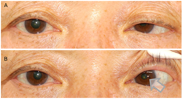
Figure 1 (A) and (B). Images of 68-year-old female who underwent endovascular treatment for left traumatic carotid-cavernous fistula 10 years ago. The patient demonstrated left-sided ptosis (A). The left eye showed miosis (B, arrow). The anisocoria increased in dim light, suggesting the left Horner syndrome.
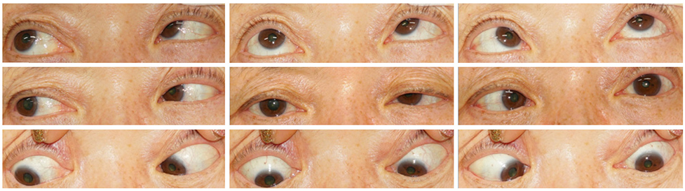
Figure 2 Images of the patient in nine diagnostic position of gaze. The patient showed 30 prism diopters left esotropia at primary gaze and −4 abduction limitation of the left eye with ptosis.
Reference
-
1. Miller NR, Subramanian PS, Patel VR. chap 18. Walsh and Hoyt's clinical neuro-ophthalmology: the essentials. 3rd ed. Philadelphia: Wolters Kluwer;2016.2. Chi CT, Nguyen D, Duc VT, et al. Direct traumatic carotid cavernous fistula: angiographic classification and treatment strategies. Study of 172 cases. Interv Neuroradiol. 2014; 20:461–475.
Article3. Gemmete JJ, Ansari SA, Gandhi DM. Endovascular techniques for treatment of carotid-cavernous fistula. J Neuroophthalmol. 2009; 29:62–71.
Article4. Liu YL, Hsieh YH, Tsai TH. Late-onset abducens nerve palsy after endovascular treatment for carotid-cavernous fistula: two case reports. Neuroophthalmology. 2014; 38:131–134.
Article5. Bou Ghannam A, Subramanian PS. Delayed onset cranial nerve palsies after endovascular coil embolization of direct carotid-cavernous fistulas. J Neuroophthalmol. 2018; 38:156–159.
Article6. Kim I, Kim JH, Kim WJ. Abducens nerve palsy caused by the ophthalmic segment of an internal carotid artery aneurysm. J Korean Ophthalmol Soc. 2018; 59:388–392.
Article7. Seo Y, Kim K, Kim WJ. Abducens nerve palsy and optic perineuritis caused by fungal sphenoidal sinusitis. J Korean Ophthalmol Soc. 2018; 59:797–801.
Article8. Kim WJ, Kim MM. Slit ventricle syndrome in pediatric patient presenting with only visual symptoms. Korean J Ophthalmol. 2017; 31:92–93.
Article9. Kang NH, Lim KH, Sung SH. Horner's syndrome with abducens nerve palsy. Korean J Ophthalmol. 2011; 25:459–462.
Article10. Kline LB, Foroozan R. Neuro-ophthalmology review manual. 7th ed. Thorofare: SLACK;2013. p. 83–122.11. American Academy of Ophthalmology. chap 10. 2018–2019 Basic and clinical science course, section 5. Neuro-ophthalmology. . San Francisco: American Academy of Ophthalmology;2018.12. Nishino K, Ito Y, Hasegawa H, et al. Cranial nerve palsy following transvenous embolization for a cavernous sinus dural arteriovenous fistula: association with the volume and location of detachable coils. J Neurosurg. 2008; 109:208–214.
Article
- Full Text Links
-
- Actions
-
Cited
- CITED
-
- Close
- Share
-
- Similar articles
-
- Carotid-cavernous Fistula Presenting as an Isolated Abducens Nerve palsy
- Isolated bilateral abducens nerve palsy due to carotid cavernous dural arteriovenous fistula
- Unilateral Abducens Nerve Palsy due to Bilateral Dural Carotid Cavernous Fistula
- Carotid Cavernous Sinus Fistula with Abducens Nerve Palsy after Le Fort I Osteotomy: A Case Report
- Isolated Abducens Nerve Palsy Caused by De Novo Pontine Cavernous Angioma
