Korean J Ophthalmol.
2011 Aug;25(4):262-267. 10.3341/kjo.2011.25.4.262.
Evaluation of the Central Macula in Commotio Retinae Not Associated with Other Types of Traumatic Retinopathy
- Affiliations
-
- 1Department of Ophthalmology, Soonchunhyang University Bucheon Hospital, Soonchunhyang University College of Medicine, Bucheon, Korea. tkpark@schmc.ac.kr
- 2Department of Ophthalmology, Kangnam Sacred Heart Hospital, Hallym University College of Medicine, Seoul, Korea.
- KMID: 1018415
- DOI: http://doi.org/10.3341/kjo.2011.25.4.262
Abstract
- PURPOSE
To report on the anatomical and functional changes to the macula in nine patients suffering from commotio retinae not accompanied by any other types of traumatic retinopathy.
METHODS
Nine injured eyes with commotio retinae were evaluated soon after ocular trauma with ophthalmic examination, including Spectral-domain optical coherence tomography (SD-OCT). In 12 eyes of 6 patients, Humphrey visual field (HVF) and multifocal electroretinogram (mfERG) were performed. Re-examinations were periodically performed for a mean of 26 days. Data from 9 injured eyes were collected and compared to data collected from the 9 non-affected eyes of the same patients.
RESULTS
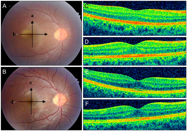
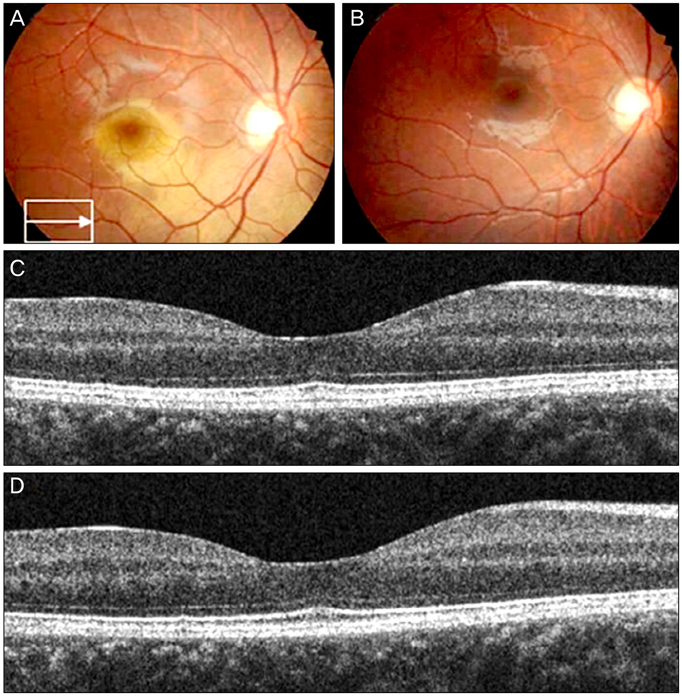
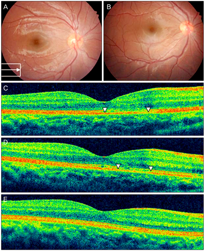
SD-OCT revealed no significant differences in the foveal thickness and total macular volume between traumatized and intact eyes in all 9 patients. Only 3 out of the 9 injured eyes showed abnormal findings in SD-OCT images such as discontinuity of the inner/outer segment (IS/OS) junction or abnormal hyper-reflectivity from the IS/OS and retinal pigment epithelium (RPE) lines in the macula. HVF and mfERG results did not show any functional deterioration in the injured eyes compared with intact eyes. During follow-up, the commotio retinae resolved in all 9 eyes. The changes to the outer retinal region detected in 3 patients by SD-OCT were also resolved.
CONCLUSIONS
Acute retinal changes in commotio retinae, not associated with other retinal pathologies, were resolved without histological and functional sequelae. In a few cases of commotio retinae, SD-OCT revealed transient abnormalities mainly observed at the IS/OS and RPE complexes.
Keyword
MeSH Terms
-
Adolescent
Adult
Child
Electroretinography
Eye Injuries/classification/*complications/pathology
Female
Follow-Up Studies
Humans
Macula Lutea/*injuries/pathology/physiopathology
Male
Middle Aged
Prognosis
Retinal Diseases/*etiology/pathology/physiopathology
Retinal Pigment Epithelium/injuries/pathology/physiopathology
Retrospective Studies
Tomography, Optical Coherence
Trauma Severity Indices
Visual Acuity
Visual Fields
Young Adult
Figure
Reference
-
1. Berlin R. Zur sogenannten commotio retinae. Klin Monatsbl Augenheilkd. 1873. 1:42–78.2. Sipperley JO, Quigley HA, Gass DM. Traumatic retinopathy in primates. The explanation of commotio retinae. Arch Ophthalmol. 1978. 96:2267–2273.3. Blight R, Hart JC. Structural changes in the outer retinal layers following blunt mechanical non-perforating trauma to the globe: an experimental study. Br J Ophthalmol. 1977. 61:573–587.4. Mansour AM, Green WR, Hogge C. Histopathology of commotio retinae. Retina. 1992. 12:24–28.5. Meyer CH, Rodrigues EB, Mennel S. Acute commotio retinae determined by cross-sectional optical coherence tomography. Eur J Ophthalmol. 2003. 13:816–818.6. Sony P, Venkatesh P, Gadaginamath S, Garg SP. Optical coherence tomography findings in commotio retina. Clin Experiment Ophthalmol. 2006. 34:621–623.7. Lai TY, Yip WW, Wong VW, Lam DS. Multifocal electroretinogram and optical coherence tomography of commotio retinae and traumatic macular hole. Eye (Lond). 2005. 19:219–221.8. Bunt-Milam AH, Black RA, Bensinger RE. Breakdown of the outer blood-retinal barrier in experimental commotio retinae. Exp Eye Res. 1986. 43:397–412.
- Full Text Links
- Actions
-
Cited
- CITED
-
- Close
- Share
- Similar articles
-
- Serial Follow-up of Commotio Retinae Using Ultra-wide Field Imaging
- A Case of the Commotio Retinae with the Myopia, Hypotony and Mydiriasis
- Utility of Ultra-wide Fundus Photography in Patients with Acute Blunt Ocular Trauma
- The Effect of Chlordiazepoxide (Olympia) on Central Serous Retinopathy
- Traumatic Optic Neuropathy Aggravated by Orbital Emphysema after Orbital Fracture


