Clin Orthop Surg.
2010 Sep;2(3):191-194. 10.4055/cios.2010.2.3.191.
Calcific Myonecrosis of the Antetibial Area
- Affiliations
-
- 1Department of Orthopaedic Surgery, Kyung Hee University East-West Neo Medical Center, Kyung Hee University School of Medicine, Seoul, Korea. mozart13@khu.ac.kr
- KMID: 999440
- DOI: http://doi.org/10.4055/cios.2010.2.3.191
Abstract
- Calcific myonecrosis is a rare late post-traumatic condition, in which a single muscle is replaced by a fusiform mass with central liquefaction and peripheral calcification. Compartment syndrome is suggested to be the underlying cause. The resulting mass may expand with time due to recurrent intralesional hemorrhage into the chronic calcified mass. A diagnosis may be difficult due to the long time between the original trauma and the symptoms of calcific myonecrosis. We encountered a 53-year-old male patient diagnosed with calcific myonecrosis in the lower leg. We report the case with a review of the relevant literature.
Keyword
MeSH Terms
Figure
-
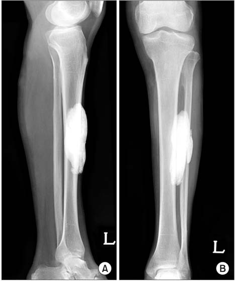
Fig. 1 Anteroposterior (A) and lateral (B) radiographs show a fusiform mass overlying the anterior compartment without an erosion of the tibia.
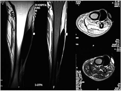
Fig. 2 Magnetic resonance images show low signal intensity in the tibialis anterior muscle.
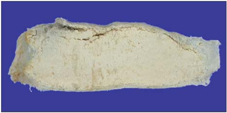
Fig. 3 The cross section of the excised mass revealing a large cystic cavity with skeletal muscle replaced by calcified material and fibrotic capsule.
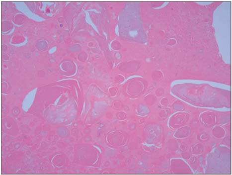
Fig. 4 Histologic examination shows an extensive amorphous pink substance with calcific material and some granular inflammatory reaction due to necrosis of the skeletal muscle and fibrin (Hematoxylin and eosin stain, ×100).
Reference
-
1. O'Keefe RJ, O'Connell JX, Temple HT, et al. Calcific myonecrosis: a late sequela to compartment syndrome of the leg. Clin Orthop Relat Res. 1995. (318):205–213.2. Holobinko JN, Damron TA, Scerpella PR, Hojnowski L. Calcific myonecrosis: keys to early recognition. Skeletal Radiol. 2003. 32(1):35–40.
Article3. Ryu KN, Bae DK, Park YK, Lee JH. Calcific tenosynovitis associated with calcific myonecrosis of the leg: imaging features. Skeletal Radiol. 1996. 25(3):273–275.
Article4. Tuncay IC, Demirors H, Isiklar ZU, Agildere M, Demirhan B, Tandogan RN. Calcific myonecrosis. Int Orthop. 1999. 23(1):68–70.
Article5. Wang JW, Chen WJ. Calcific myonecrosis of the leg: a case report and review of the literature. Clin Orthop Relat Res. 2001. (389):185–190.6. Larson RC, Sierra RJ, Sundaram M, Inwards C, Scully SP. Calcific myonecrosis: a unique presentation in the upper extremity. Skeletal Radiol. 2004. 33(5):306–309.
Article7. Zohman GL, Pierce J, Chapman MW, Greenspan A, Gandour-Edwards R. Calcific myonecrosis mimicking an invasive soft-tissue neoplasm: a case report and review of the literature. J Bone Joint Surg Am. 1998. 80(8):1193–1197.8. Early JS, Ricketts DS, Hansen ST. Treatment of compartmental liquefaction as a late sequelae of a lower limb compartment syndrome. J Orthop Trauma. 1994. 8(5):445–448.
Article9. Malisano LP, Hunter GA. Liquefaction and calcification of a chronic compartment syndrome of the lower limb. J Orthop Trauma. 1992. 6(2):245–247.
Article10. Viau MR, Pedersen HE, Salciccioli GG, Manoli A 2nd. Ectopic calcification as a late sequela of compartment syndrome: report of two cases. Clin Orthop Relat Res. 1983. (176):178–180.
