Calcific Myonecrosis of the Lower Extremities: A Case Report
- Affiliations
-
- 1Department of Radiology, Seoul Veterans Hospital, Korea. orabykim@paran.com
- 2Department of Pathology, Seoul Veterans Hospital, Korea.
- KMID: 1443585
- DOI: http://doi.org/10.3348/jksr.2011.64.1.71
Abstract
- Calcific myonecrosis is a rare and latent condition of the lower extremities after a trauma and is characterized by the formation of a fusiform mass lesion in the anterior compartment of the leg showing peripheral dystrophic calcification and central liquefaction. We report the radiologic findings of calcific myonecrosis in a patient with a lower extremity calcified mass lesion.
MeSH Terms
Figure
-
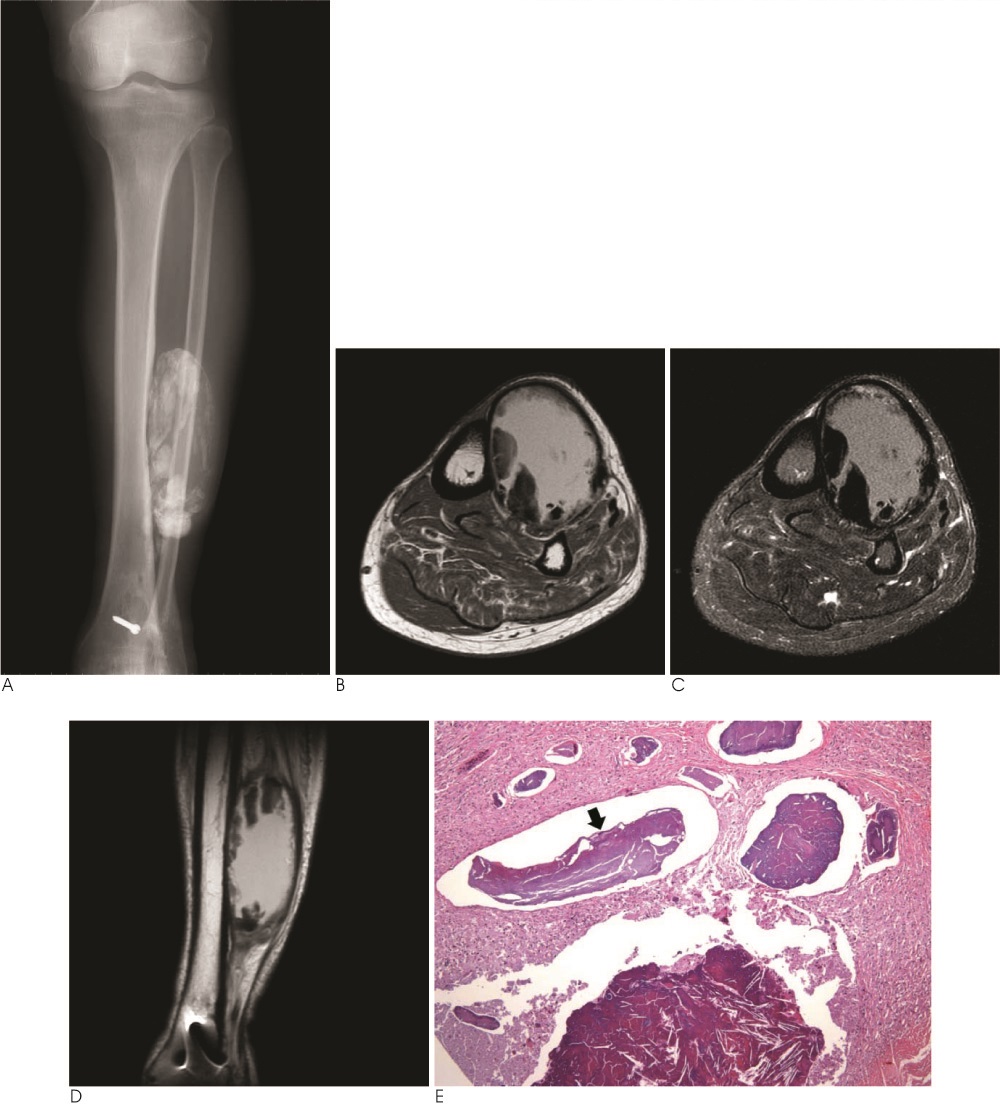
Fig. 1 A 73-year-old man with calcific myonecrosis presented as a painless, slowly enlarging mass on the anterolateral aspect of left leg. A. Anteroposterior radiograph shows lobulated, fusiform soft tissue mass in the anterior compartment of the left leg with well defined peripheral plaque-like calcifications and adjacent tibial cortex erosion. B. Axial T1-weighted MR image shows a thick and nodular low signal intensity peripheral rim corresponding to the distribution of calcification. The central portion has a homogenous and high signal intensity. C. Axial fat suppressed T2-weighted MR image shows the heterogenous calcified mass in the anterior compartment of the leg with a predominant high signal intensity central cystic appearance. D. A coronal proton density-weighted MR image shows the extent of the soft tissue mass in the lower leg. E. Photomicrograph of the histopathologic specimen shows a characteristic histiocytic reaction with dystrophic calcification (arrow) (H & E stain, ×100).
Reference
-
1. Holobinko JN, Damron TA, Scerpella PR, Hojnowski L. Calcific myonecrosis: keys to early recognition. Skeletal Radiol. 2003; 32:35–40.2. Muramatsu K, Ihara K, Seki T, Imagama T, Taguchi T. Calcific myonecrosis of the lower leg: diagnosis and options of treatment. Arch Orthop Trauma Surg. 2009; 129:935–939.3. Larson RC, Sierra RJ, Sundaram M, Inwards C, Scully SP. Calcific myonecrosis: a unique presentation in the upper extremity. Skeletal Radiol. 2004; 33:306–309.4. O'Dwyer HM, Al-Nakshabandi NA, Al-Muzahmi K, Ryan A, O'Connell JX, Munk PL. Calcific myonecrosis: keys to recognition and management. AJR Am J Roentgenol. 2006; 187:W67–W76.5. Tuncay IC, Demirors H, Isiklar ZU, Agildere M, Demirhan B, Tandogan RN. Calcific myonecrosis. Int Orthop. 1999; 23:68–70.6. Janzen DL, Connell DG, Vaisler BJ. Calcific myonecrosis of the calf manifesting as an enlarging soft-tissue mass: imaging features. AJR Am J Roentgenol. 1993; 160:1072–1074.7. Dhillon M, Davies AM, Benham J, Evans N, Mangham DC, Grimer RJ. Calcific myonecrosis: a report of ten new cases with an emphasis on MR imaging. Eur Radiol. 2004; 14:1974–1979.8. Viau MR, Pedersen HE, Salciccioli GG, Manoli A 2nd. Ectopic calcification as a late sequela of compartment syndrome. Report of two cases. Clin Orthop Relat Res. 1983; 178–180.9. Snyder BJ, Oliva A, Buncke HJ. Calcific myonecrosis following compartment syndrome: report of two cases, review of the literature, and recommendations for treatment. J Trauma. 1995; 39:792–795.10. Wang JW, Chen WJ. Calcific myonecrosis of the leg: a case report and review of the literature. Clin Orthop Relat Res. 2001; 185–190.
- Full Text Links
-
- Actions
-
Cited
- CITED
-
- Close
- Share
-
- Similar articles
-
- Calcific Myonecrosis of the Calf
- Calcific Myonecrosis of the Antetibial Area
- Extensive calcific myonecrosis of the lower leg treated with free tissue transfer
- Thoracolumbar Paraspinal Myonecrosis after Aortic Dissection
- Arthroscopic treatment of chronic calcific tendinitis with intraosseous migration: a case report
