The Surgical Treatment for Spinal Intradural Extramedullary Tumors
- Affiliations
-
- 1Department of Orthopedic Surgery, Seoul Sacred Heart General Hospital, Seoul, Korea. hoon0319@gamil.com
- KMID: 999396
- DOI: http://doi.org/10.4055/cios.2009.1.3.165
Abstract
- BACKGROUND
We wanted to investigate the results of surgical treatment and analyze the factors that have an influence on the neurologic symptoms and prognosis of spinal intradural extramedullary (IDEM) tumors. METHODS: The spinal IDEM tumor patients (11 cases) who had been treated by surgical excision and who were followed up more than 1 year were retrospectively analyzed. Pain was evaluated by the visual analogue scale (VAS) and the neurologic function was assessed by Nurick's grade. The pathological diagnosis, the preoperative symptom duration, the tumor location on the sagittal and axial planes and the percentage of tumor occupying the intradural space were investigated. In addition, all these factors were analyzed in relation to the degree of the preoperative symptoms and the prognosis. On the last follow-up, the MRI was checked to evaluate whether or not the tumor had recurred. RESULTS: The most common diagnosis was schwannomas (73%), followed by meningiomas (18%). The percentage of tumor occupying the intradural space was 82.9 +/- 9.4%. The VAS score was reduced in all cases from 8.0 +/- 1.2 to 1.2 +/- 0.8 (p = 0.003) and the Nurick's grade was improved in all cases from 3.0 +/- 1.3 to 1.0 +/- 0.0 (p = 0.005). The preoperative symptoms were correlated with only the percentage of tumor occupying the intradural space (VAS; r2 = 0.75, p = 0.010, Nurick's grade; r2 = 0.69, p = 0.019). One case of schwannoma recurred. CONCLUSIONS: The degree of neurologic symptoms was correlated with the percentage of tumor occupying the intradural space. All the tumors were able to be excised through the posterior approach. The postoperative neurologic recovery was excellent in all the cases regardless of any condition. Therefore, aggressive surgical excision is recommended even for cases with a long duration of symptoms or a severe neurologic deficit.
MeSH Terms
Figure
-
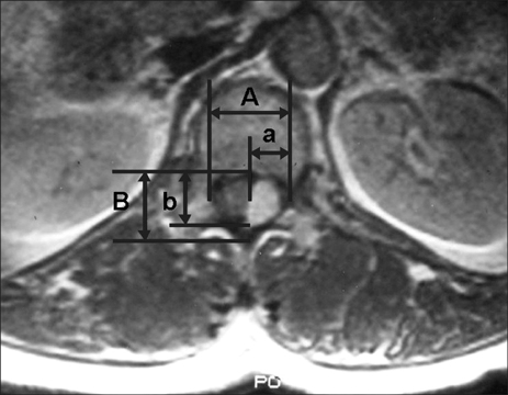
Fig. 1 This picture shows how to calculate the percentage of tumor occupying the intradural space on an axial MRI film. It is as follows: {(a + b) / (A + B)} × 100. A: transverse diameter of the intradural space, B: longitudinal diameter of the intradural space, a: transverse diameter of the tumor mass, b: longitudinal diameter of the tumor mass.

Fig. 2 This shows a nerve fiber over the surface of schwannoma. It was separable from the tumor mass.
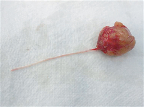
Fig. 3 This shows a nerve fiber that penetrated a schwannoma. But any distal connected fiber was not identified.
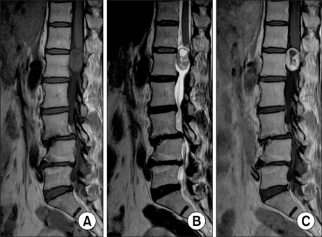
Fig. 4 Schawannoma: (A) The T1 weighted image shows signal intensity that is similar to that of the spinal cord. (B) The T2 weighted image shows ir -regular and higher signal in tensity than that of the CSF. (C) The contrast-enhanced MR often shows an irregular margin and ring-shape enhancement.

Fig. 5 Meningioma: (A, B) The T1 & T2 weighted images both show slightly lower intense than that of the cord and this is a homogenous lesion. (C) Contrast-enhanced MR shows high homogenous signal intensity of tumor.
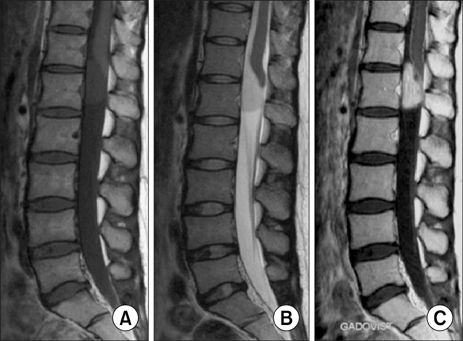
Fig. 6 Ependymoma: (A) The T1 weighted image shows signal intensity that is similar to the spinal cord. (B) The T2 weighted image shows higher signal intensity than that of the spinal cord. (C) Contrast-enhanced MR shows a well defined margin and regular intensity lesion.
Reference
-
1. Albanese V, Platania N. Spinal intradural extramedullary tumors: personal experience. J Neurosurg Sci. 2002. 46(1):18–24.2. el-Mahdy W, Kane PJ, Powell MP, Crockard HA. Spinal intradural tumours: Part I Extramedullary. Br J Neurosurg. 1999. 13(6):550–557.3. Tredway TL, Santiago P, Hrubes MR, Song JK, Christie SD, Fessler RG. Minimally invasive resection of intradural-extramedullary spinal neoplasms. Neurosurgery. 2006. 58(1):Suppl. ONS52–ONS58.
Article4. Nurick S. The natural history and the results of surgical treatment of the spinal cord disorder associated with cervical spondylosis. Brain. 1972. 95(1):101–108.
Article5. Helseth A, Mork SJ. Primary intraspinal neoplasms in Norway, 1955 to 1986: a population-based survey of 467 patients. J Neurosurg. 1989. 71(6):842–845.6. Solero CL, Fornari M, Giombini S, et al. Spinal meningiomas: review of 174 operated cases. Neurosurgery. 1989. 25(2):153–160.
Article7. Saito T, Arizono T, Maeda T, Terada K, Iwamoto Y. A novel technique for surgical resection of spinal meningioma. Spine. 2001. 26(16):1805–1808.
Article8. Ahn DK, Jung KW, Lee S, Choi DJ, Park JS, Cha SG. A 6-year observation of multiple spinal schwannomas before excision: 1 case report. J Korean Soc Spine Surg. 2003. 10(3):277–282.
Article9. Asazuma T, Toyama Y, Watanabe M, Suzuki N, Fujimura Y, Hirabayashi K. Clinical features associated with recurrence of tumours of the spinal cord and cauda equina. Spinal Cord. 2003. 41(2):85–89.
Article10. Parsa AT, Lee J, Parney IF, Weinstein P, McCormick PC, Ames C. Spinal cord and intradural-extraparenchymal spinal tumors: current best care practices and strategies. J Neurooncol. 2004. 69(1-3):291–318.
Article11. Prevedello DM, Koerbel A, Tatsui CE, et al. Prognostic factors in the treatment of the intradural extramedullary tumors: a study of 44 cases. Arq Neuropsiquiatr. 2003. 61(2A):241–247.12. Kim P, Ebersold MJ, Onofrio BM, Quast LM. Surgery of spinal nerve schwannoma: risk of neurological deficit after resection of involved root. J Neurosurg. 1989. 71(6):810–814.13. Gottfried ON, Gluf W, Quinones-Hinojosa A, Kan P, Schmidt MH. Spinal meningiomas: surgical management and outcome. Neurosurg Focus. 2003. 14(6):e2.
Article14. Slin'ko EI, Al-Qashqish II. Intradural ventral and ventrolateral tumors of the spinal cord: surgical treatment and results. Neurosurg Focus. 2004. 17(1):ECP2.15. Rezai AR, Woo HH, Lee M, Cohen H, Zagzag D, Epstein FJ. Disseminated ependymomas of the central nervous system. J Neurosurg. 1996. 85(4):618–624.
Article16. Klekamp J, Samii M. Surgical results of spinal meningiomas. Acta Neurochir Suppl. 1996. 65:77–81.
Article17. Gezen F, Kahraman S, Canakci Z, Beduk A. Review of 36 cases of spinal cord meningioma. Spine. 2000. 25(6):727–731.
Article18. Shin BJ, Lee JC, Yoon TK, et al. Surgical treatments of intradural extramedullary tumor. J Korean Soc Spine Surg. 2002. 9(3):230–237.
Article
- Full Text Links
-
- Actions
-
Cited
- CITED
-
- Close
- Share
-
- Similar articles
-
- Removal of Intradural-Extramedullary Spinal Cord Tumors with Unilateral Limited Laminectomy
- Non-Enhancing Intradural Extramedullary Ependymoma: A Case Report
- Intradural Extramedullary Spinal Ependymoma: A Case Report of Malignant Transformation Occurring
- MR Imaging of Intradural Extramedullary Tuberculoma of the Spinal Cord: Report of Two Cases
- Exploring Unilateral Biportal Endoscopy for Lumbar Intradural Lesions: A Technical and Video Report on Benefits and Key Considerations
