A Case of Histiocytoid Variant Eccrine Sweat Gland Carcinoma of the Orbit
- Affiliations
-
- 1Department of Ophthalmology, Seoul Veterans Hospital, Seoul, Korea. ezer75@hanmail.net
- 2Department of Pathology, Kangnam Sacred Heart Hospital, Hallym University College of Medicine, Seoul, Korea.
- KMID: 994413
- DOI: http://doi.org/10.3341/kjo.2011.25.1.54
Abstract
- A 79-year-old male presented with left ocular pain. Evisceration and silicone ball implantation were performed after a diagnosis of phthisis. He returned six weeks later because of left facial erythematous swelling, tenderness, mild fever, chills and cough. His condition was diagnosed as orbital cellulitis. Despite two weeks of empirical antibiotic therapy, the symptoms worsened. A subsequent orbital computed tomography scan revealed enhanced soft tissue infiltrations in his left orbit and eyelid. Biopsy showed a diffusely infiltrating tumor of signet ring cell cytology. A systemic evaluation revealed multiple bone metastases. Based on this evidence, the patient was diagnosed with a very rare case of histiocytoid variant eccrine sweat gland carcinoma with multiple bone metastases.
MeSH Terms
Figure
-
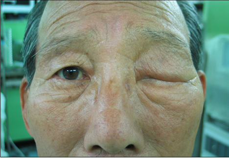
Fig. 1 Clinical photograph of the erythematous swelling of the left eyelid.
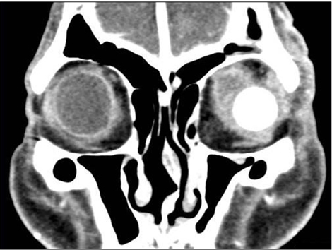
Fig. 2 Enhanced extraconal and intraconal infiltrations in the left orbit, and an inferolateral view of the silicone ball sphere in the eyelid.
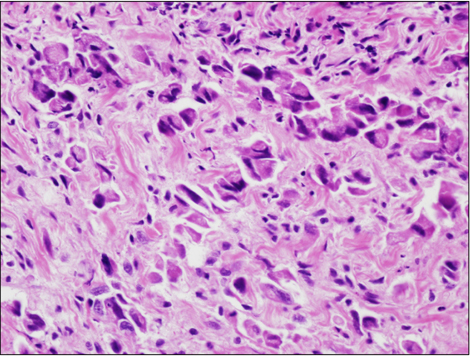
Fig. 3 The tumors were composed of cells with hyperchromatic eccentric nuclei (suggesting signet ring cells), prominent eosinophilic cytoplasm, and intracytoplasmic vacuolations (H&E, ×400).
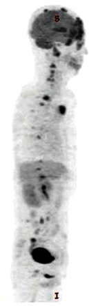
Fig. 4 A positron emission tomography image revealed the multiple intense hypermetabolic foci on the left orbit and the skull as well as along the spine.
Cited by 1 articles
-
A Case of Primary Signet Ring Cell Carcinoma of the Lower Eyelid
Seon Ae Shin, Sang Duck Kim, Ki Jung Yun
J Korean Ophthalmol Soc. 2014;55(4):611-615. doi: 10.3341/jkos.2014.55.4.611.
Reference
-
1. Mortensen AL, Heegaard S, Clemmensen O, Prause JU. Signet ring cell carcinoma of the eyelid - the monocle tumour. APMIS. 2008. 116:326–332.2. Langel DJ, Yeatts RP, White WL. Primary signet ring cell carcinoma of the eyelid: report of a case demonstrating further analogy to lobular carcinoma of the breast with a literature review. Am J Dermatopathol. 2001. 23:444–449.3. Kramer TR, Grossniklaus HE, McLean IW, et al. Histiocytoid variant of eccrine sweat gland carcinoma of the eyelid and orbit: report of five cases. Ophthalmology. 2002. 109:553–559.4. Swinson B, Ryan F, Barrett AW, et al. Histiocytoid eccrine sweat gland carcinoma of the eyelid: report of a case. Clin Exp Dermatol. 2006. 31:786–789.5. Hoppenreijs VP, Reuser TT, Mooy CM, et al. Syringomatous carcinoma of the eyelid and orbit: a clinical and histopathological challenge. Br J Ophthalmol. 1997. 81:668–672.6. Krishnakumar S, Mohan ER, Babu K, et al. Eccrine duct carcinoma of the eyelid mimicking meibomian carcinoma: clinicopathological study of a case. Surv Ophthalmol. 2003. 48:439–446.7. Jakobiec FA, Austin P, Iwamoto T, et al. Primary infiltrating signet ring carcinoma of the eyelids. Ophthalmology. 1983. 90:291–299.8. Wollensak G, Witschel H, Böhm N. Signet ring cell carcinoma of the eccrine sweat glands in the eyelid. Ophthalmology. 1996. 103:1788–1793.
