Korean J Gastroenterol.
2010 Jun;55(6):394-398. 10.4166/kjg.2010.55.6.394.
A Case of Large Retroperitoneal Lipoma Mimicking Liposarcoma
- Affiliations
-
- 1Department of Internal Medicine, Chonnam National University Medical School, Gwangju, Korea. yejoo@chonnam.ac.kr
- 2Department of General Surgery, Chonnam National University Medical School, Gwangju, Korea.
- KMID: 948769
- DOI: http://doi.org/10.4166/kjg.2010.55.6.394
Abstract
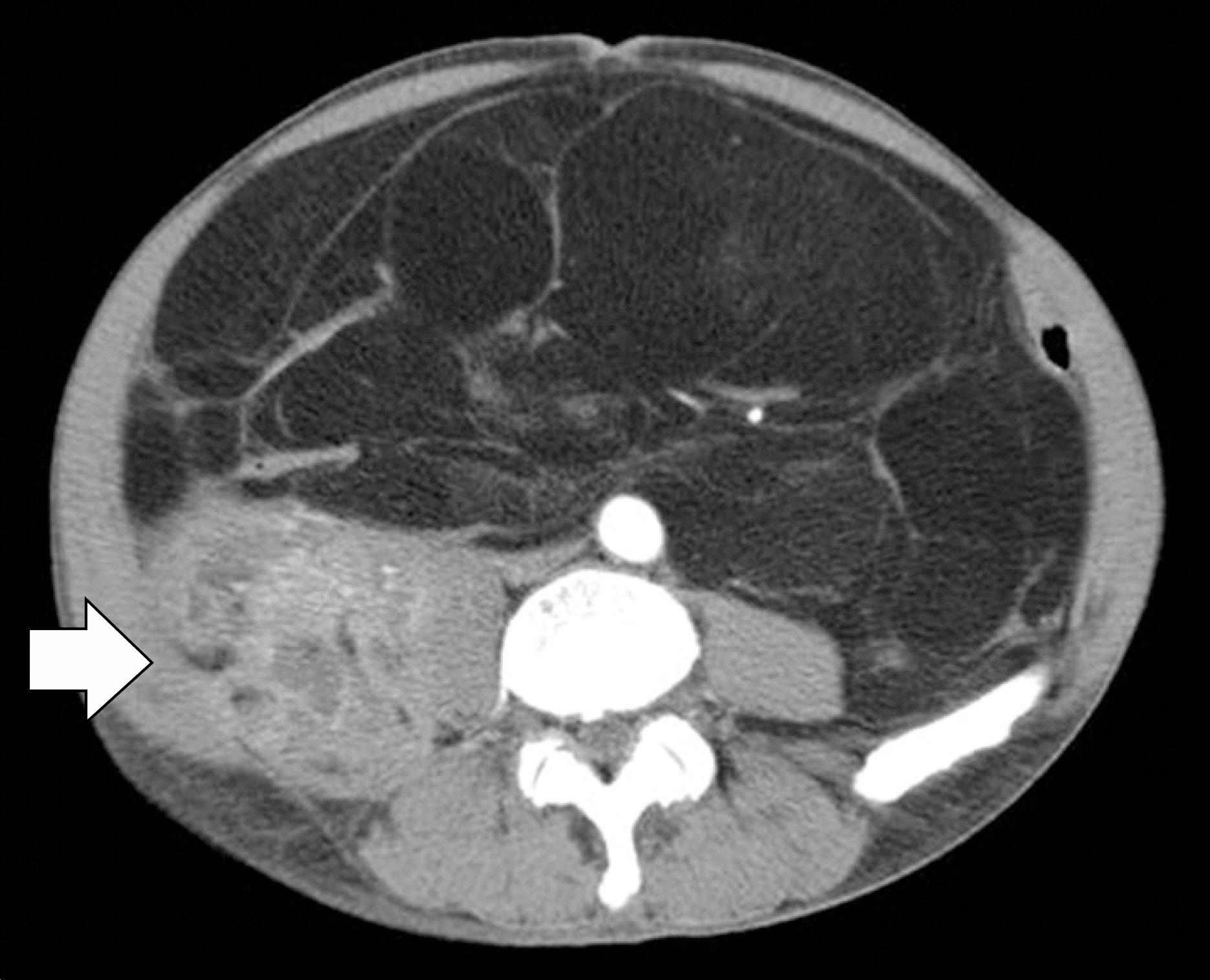
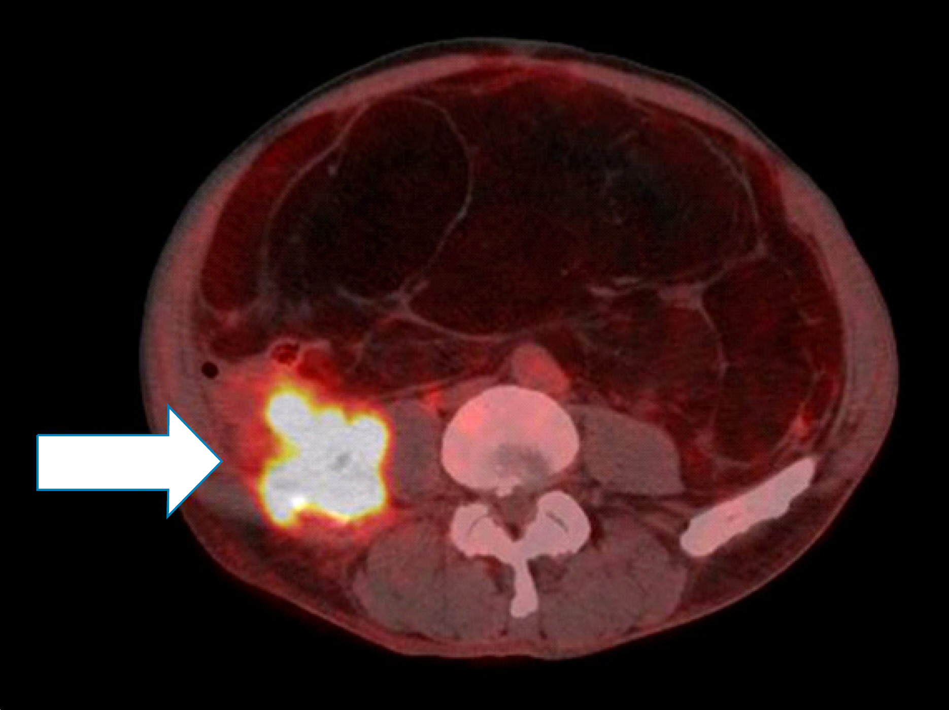
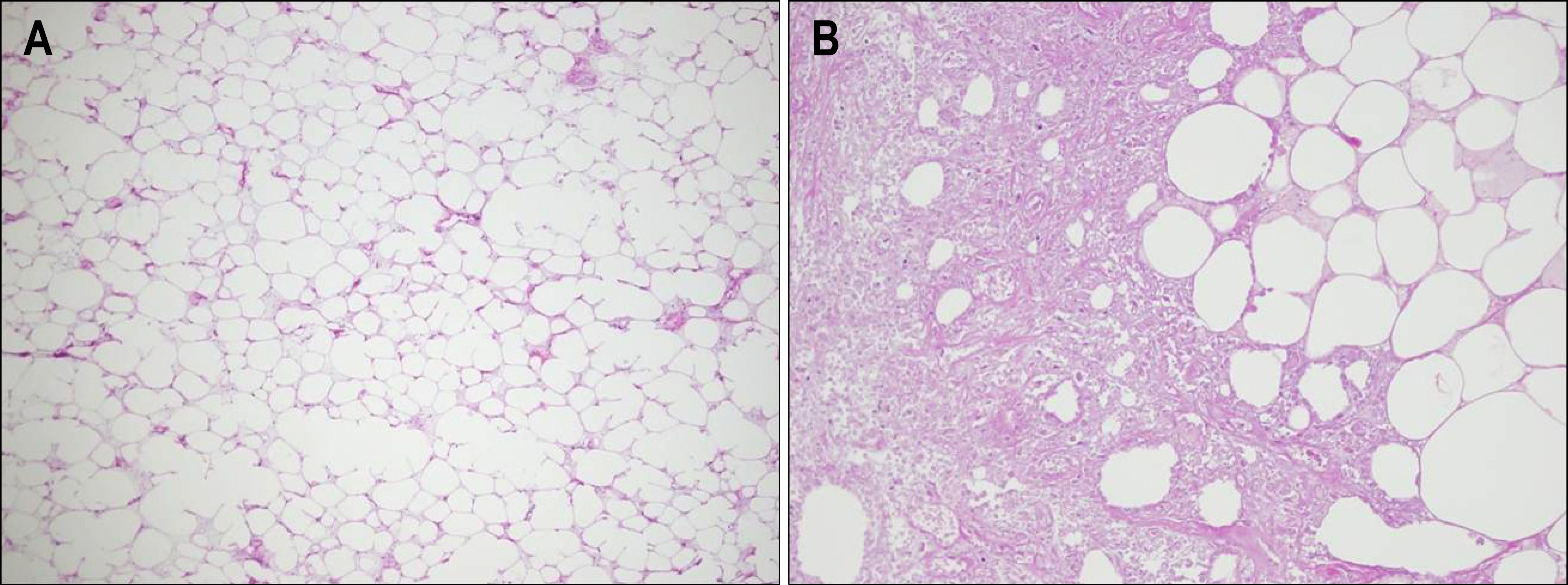
- Lipomas are the most common benign tumors of adipose tissue among adults. Lipomas can occur almost anywhere in the trunk, extremities, mediastinum, and pelvis, but retroperitoneal lipomas are extremely rare. It should be distinguished from well differentiated liposarcoma in order to provide the appropriate treatment and follow up. We experienced a case of 60-year-old patient with large retroperitoneal lipoma mimicking liposarcoma causing palpable abdominal mass and pain. Abdominal computerized tomography (CT) showed 33x22 cm sized bulky fat-containing mass with contrast enhanced solid portion in right retroperitoneum. Positron emission tomograpgy (PET) revealed increased 18F-FDG uptake at solid portion shown in abdominal CT. Imaging studies confirmed a high index of suspicion on liposarcoma. Laparotomy showed a large encapsulating tumor arising from retroperitoneum with fat necrosis. Pathologic examination of resected specimen revealed normal mature adipocytes without atypical cells, compatible with lipoma.
Keyword
MeSH Terms
Figure
Reference
-
1. Martinez CA, Palma RT, Waisberg J. Giant retroperitoneal lipoma: a case report. Arq Gastroenterol. 2003; 40:251–255.
Article2. OcKuly EA. Retroperitoneal perirenal lipoma. J Urol. 1937; 37:619–630.3. Peitsidis P, Peitsidou A, Tsekoura V, Zervoudis S, Akrivos T. Managment of large retroperitoneal lipoma in a 12-year-old patient. Urology. 2009; 73:797–799.4. Pemberton JJ, Whitlock ME. Large retroperitoneal lipoma. Report of case. Surg Clin North Am. 1934; 14:601–606.5. Seo IJ, Lee JK, Park SY, Jeon SH, Seong IG, Han BH. A case of huge retroperitoneal lipoma. Korean J Urol. 1996; 7:824–828.6. Singaporewalla RM, Thamboo TP, Rauff A, Cheah WK, Mukherjee JJ. Acute abdominal pain secondary to retroperitoneal bleeding from a giant adrenal lipoma with review of literature. Asian J Surg. 2009; 32:172–176.
Article7. Foa C, Mainquene C, Dupre F, et al. Rearrangement involving chromosomes 1 and 8 in a retroperitoneal lipoma. Cancer Genet Cytogenet. 2002; 133:156–159.
Article8. Amstrong JR, Cohn I Jr. Primary malignant retroperitoneal tumors. Am J Surg. 1965; 110:937–943.9. Munk PL, Lee MJ, Janzen DL, et al. Lipoma and liposarcoma: evaluation using CT and MR imaging. AJR Am J Roentgenol. 1997; 169:589–594.
Article10. Craig WD, Fanburg-Smith JC, Henry LR, Guerrero R, Barton JH. Fat-containing lesions of the retroperitoneum: radio-logic-pathologic correlation. Radiographics. 2009; 29:261–290.
Article11. Takahashi Y, Irisawa A, Bhutani MS, et al. Two cases of retroperitoneal liposarcoma diagnosed using endoscopic ultrasound-guided fine-needle aspiration (EUS-FNA). Diagn Ther Endosc. 2009; 2009:673194.
Article12. Nemanqani D, Mourad WA. Cytomorphologic features of fine-needle aspiration of liposarcoma. Diagn Cytopathol. 1999; 20:67–69.
Article13. Laurino L, Furlanetto A, Orvieto E, Del Tos AP. Well-differ-entiated liposarcoma (atypical lipomatous tumours). Semin Diagn Pathol. 2001; 18:258–262.14. Oh BR, Sung JS, Chung SY, Choi SN. The expression Ki-67 and p53 protein in intraabdominal liposarcomas. J Korean Surg Soc. 2004; 66:333–337.15. Sato T, Nishimura G, Nonomura A, Miwa K. Intraabdominal and retroperitoneal liposarcomas. Int Surg. 1999; 84:163–167.16. Takao H, Yamahira K, Watanabe T. Encapsulated fat necrosis mimicking abdominal liposarcoma: computed tomography findings. J Comput Assist Tomogr. 2004; 28:193–194.17. Kinne DW, Chu FC, Huvos AG, Yagoda A, Fortner JG. Treatment of primary and recurrent retroperitoneal liposarcoma. Twenty-five-year experience at Memorial Hospital. Cancer. 1973; 31:53–64.18. Clark MA, Fisher C, Judson I, Thomas JM. Soft-tissue sarco-mas in adults. N Engl J Med. 2005; 353:701–711.
Article


