Evaluation of Left Atrial Volumes Using Multidetector Computed Tomography: Comparison with Echocardiography
- Affiliations
-
- 1Department of Radiology and Research Institute of Radiological Science, Yonsei University Health System, Yonsei University College of Medicine, Gangnam Severance Hospital, Seoul 135-720, Korea. thkim1@yuhs.ac
- 2Department of Cardiology, Yonsei University Health System, Yonsei University College of Medicine, Gangnam Severance Hospital, Seoul 135-720, Korea.
- KMID: 946268
- DOI: http://doi.org/10.3348/kjr.2010.11.3.286
Abstract
OBJECTIVE
To prospectively assess the relationship between the two different measurement methods for the evaluation of left atrial (LA) volume using cardiac multidetector computed tomography (MDCT) and to compare the results between cardiac MDCT and echocardiography. MATERIALS AND METHODS: Thirty-five patients (20 men, 15 women; mean age, 60 years) underwent cardiac MDCT angiography for coronary artery disease. The LA volumes were measured using two different methods: the two dimensional (2D) length-based (LB) method measured along the three-orthogonal planes of the LA and the 3D volumetric threshold-based (VTB) method measured according to the threshold 3D segmentation of the LA. The results obtained by cardiac MDCT were compared with those obtained by echocardiography. RESULTS: The LA end-systolic and end-diastolic volumes (LAESV and LAEDV) measured by the 2D-LB method correlated well with those measured by the 3D-VTB method using cardiac MDCT (r = 0.763, r = 0.786, p = 0.001). However, there was a significant difference in the LAESVs between the two measurement methods using cardiac MDCT (p < 0.05). The LAESV measured by cardiac MDCT correlated well with measurements by echocardiography (r = 0.864, p = 0.001), however with a significant difference (p < 0.01) in their volumes. The cardiac MDCT overestimated the LAESV by 22% compared to measurements by echocardiography. CONCLUSION: A significant correlation was found between the two different measurement methods for evaluating LA volumes by cardiac MDCT. Further, cardiac MDCT correlates well with echocardiography in evaluating the LA volume. However, there are significant differences in the LAESV between the two measurement methods using cardiac MDCT and between cardiac MDCT and echocardiography.
Keyword
MeSH Terms
-
Adult
Aged
Aged, 80 and over
Atrial Function, Left
*Cardiac Volume
Contrast Media/diagnostic use
Coronary Artery Disease/*radiography/ultrasonography
Electrocardiography/methods
Female
Heart Atria/radiography/ultrasonography
Humans
Imaging, Three-Dimensional/methods
Male
Middle Aged
Observer Variation
Prospective Studies
Radiographic Image Enhancement/methods
Tomography, X-Ray Computed/*methods
Triiodobenzoic Acids/diagnostic use
Figure
-
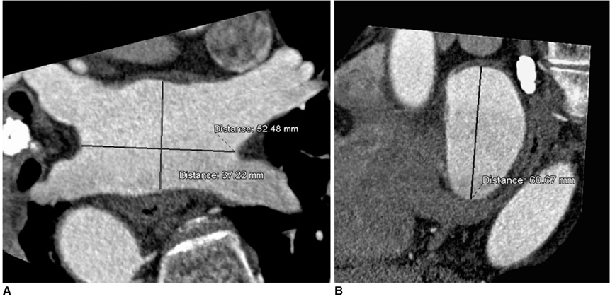
Fig. 1 Measurement of left atrial size using 2D length-based method of cardiac multidetector CT. A. Oblique axial image of left atrium. Transverse diameter of left atrium was measured at distance between right and left pulmonary veins. Anterior-posterior diameter of left atrium was measured at midpoint of transverse diameter. B. Sagittal view of left atrium. Longitudinal diameter of left atrium was measured at midpoint of transverse diameter.
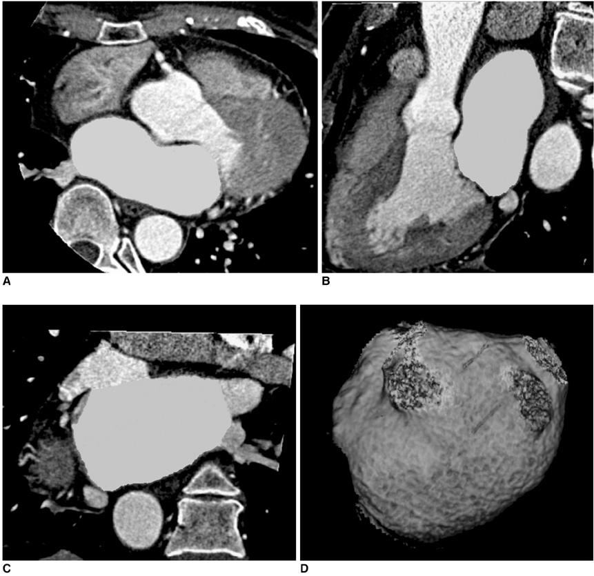
Fig. 2 Measurement of left atrial volume using 3D volume threshold-based method of cardiac multidetector CT. A-C. Axial, sagittal, and coronal views of left atrium. Endocardial contours of left atrium were traced on axial slices. Lowest value of CT attenuation was applied to cover contrast-enhanced whole left atrial cavity within region of interest. Included left atrial volume was confirmed by CT attenuation in three-orthogonal planes. D. Volume-rendering threshold image of left atrium. Pulmonary vein confluences and atrial appendage were excluded from left atrial volume measurement.
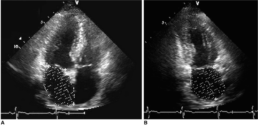
Fig. 3 Measurement of left atrial volume using modified biplane Simpson's rule method by echocardiography. A, B. Apical four-chamber and apical two-chamber views at ventricular end-systole for measurement of maximum left atrial volume.
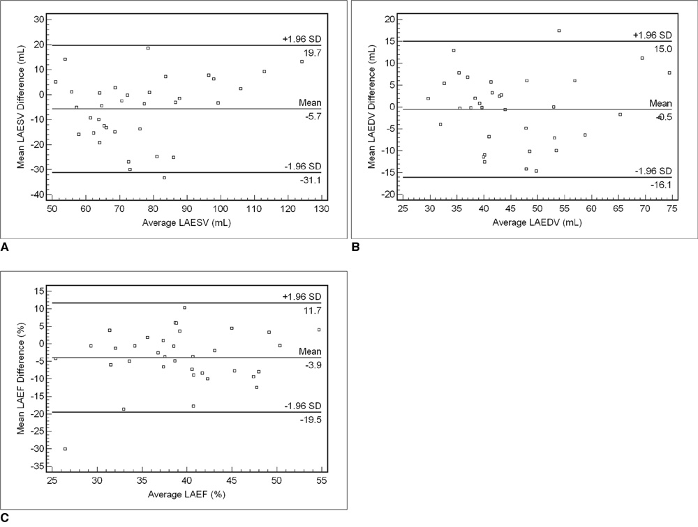
Fig. 4 Bland-Altman plots for determining left atrial volumes and function between two measurement methods of cardiac multidetector CT. A-C. Plots show relationships between 2D length-based method (2D LBM) and 3D volumetric threshold-based method (3D VTBM) of cardiac multidetector CT for measurement of left atrial end-systolic volume (A), left atrial end-diastolic volume (B), and left atrial ejection fraction (C). Mean differences (y-axes) between each pair ([mean 2D LBM] - [mean 3D VTBM]) are plotted against average values (x-axes) of same pair ([{mean 2D LBM} + {mean 3D VTBM}]/2). The mean differences between the two measurement methods by cardiac multidetector CT were significant for left atrial end-systolic volume and left atrial ejection fraction (p < 0.05), but not for left atrial end-diastolic volume (p > 0.05). LAESV = left atrial end-systolic volume, LAEDV = left atrial end-diastolic volume, LAEF = left atrial ejection fraction

Fig. 5 Linear regression analysis and Bland-Altman plots for left atrial end-systolic volume between cardiac multidetector CT and echocardiography. LAESV = left atrial end-systolic volume, ECHO = echocardiography A. Left atrial end-systolic volumes were plotted by linear regression for cardiac multidetector CT with 3D volumetric threshold-based method (3D VTBM) and echocardiography. Slope, correlation coefficient, and p value were 0.949, 0.864, and less than 0.001 (Y = 20.536 + 0.949X, r = 0.864), respectively. B. Bland-Altman plots showing relationship between cardiac multidetector CT with 3D volumetric threshold-based method and echocardiography with modified biplane Simpson's method (MBSM) for left atrial end-systolic volume. Mean differences (y-axes) between each pair ([mean 3D VTBM] - [mean MBSM]) are plotted against average values (x-axes) of same pair ([{mean 3D VTBM} + {mean MBSM}]/2). Results showed that echocardiography underestimated left atrial end-systolic volume by 22% compared to cardiac multidetector CT (p < 0.05).
Cited by 1 articles
-
Relationship between Epicardial Fat Accumulation and Left Atrial Reverse Remodeling after Catheter Ablation of Atrial Fibrillation
Jeong Yoon Lee, Yu-Whan Oh, Young-Hoon Kim, Jaemin Shim, Sung Ho Hwang
J Korean Soc Radiol. 2019;80(5):930-941. doi: 10.3348/jksr.2019.80.5.930.
Reference
-
1. Beinart R, Boyko V, Schwammenthal E, Kuperstein R, Sagie A, Hod H, et al. Long-term prognostic significance of left atrial volume in acute myocardial infarction. J Am Coll Cardiol. 2004. 44:327–334.2. Kizer JR, Bella JN, Palmieri V, Liu JE, Best LG, Lee ET, et al. Left atrial diameter as an independent predictor of first clinical cardiovascular events in middle-aged and elderly adults: the Strong Heart Study (SHS). Am Heart J. 2006. 151:412–418.3. Rossi A, Cicoira M, Zanolla L, Sandrini R, Golia G, Zardini P, et al. Determinants and prognostic value of left atrial volume in patients with dilated cardiomyopathy. J Am Coll Cardiol. 2002. 40:1425.4. Tsang TS, Barnes ME, Bailey KR, Leibson CL, Montgomery SC, Takemoto Y, et al. Left atrial volume: important risk marker of incident atrial fibrillation in 1655 older men and women. Mayo Clin Proc. 2001. 76:467–475.5. Feinberg MS, Waggoner AD, Kater KM, Cox JL, Lindsay BD, Perez JE. Restoration of atrial function after the maze procedure for patients with atrial fibrillation. Assessment by Doppler echocardiography. Circulation. 1994. 90:II285–II292.6. Yashima N, Nasu M, Kawazoe K, Hiramori K. Serial evaluation of atrial function by Doppler echocardiography after the maze procedure for chronic atrial fibrillation. Eur Heart J. 1997. 18:496–502.7. Jessurun ER, van Hemel NM, Kelder JC, Defauw JA, Brutel de la Rivière A, Ernst JM, et al. The effect of maze operations on atrial volume. Ann Thorac Surg. 2003. 75:51–56.8. Bartunek J, Vantrimpont PJ, De Bruyne B. Left atrial-volume determination by echocardiography. Validation by biplane angiography in the setting of mitral balloon valvuloplasty. Int J Card Imaging. 1994. 10:263–268.9. Rodevan O, Bjornerheim R, Ljosland M, Maehle J, Smith HJ, Ihlen H. Left atrial volumes assessed by three- and two-dimensional echocardiography compared to MRI estimates. Int J Card Imaging. 1999. 15:397–410.10. Kircher B, Abbott JA, Pau S, Gould RG, Himelman RB, Higgins CB, et al. Left atrial volume determination by biplane two-dimensional echocardiography: validation by cine computed tomography. Am Heart J. 1991. 121:864–871.11. Christiaens L, Lequeux B, Ardilouze P, Ragot S, Mergy J, Herpin D, et al. A new method for measurement of left atrial volumes using 64-slice spiral computed tomography: comparison with two-dimensional echocardiographic techniques. Int J Cardiol. 2009. 131:217–224.12. Achenbach S, Ulzheimer S, Baum U, Kachelriess M, Ropers D, Giesler T, et al. Noninvasive coronary angiography by retrospectively ECG-gated multislice spiral CT. Circulation. 2000. 102:2823–2828.13. Raff GL, Gallagher MJ, O'Neill WW, Goldstein JA. Diagnostic accuracy of noninvasive coronary angiography using 64-slice spiral computed tomography. J Am Coll Cardiol. 2005. 46:552–557.14. Mollet NR, Cademartiri F, van Mieghem CA, Runza G, McFadden EP, Baks T, et al. High-resolution spiral computed tomography coronary angiography in patients referred for diagnostic conventional coronary angiography. Circulation. 2005. 112:2318–2323.15. Kim TH, Hur J, Kim SJ, Kim HS, Choi BW, Choe KO, et al. Two-phase reconstruction for the assessment of left ventricular volume and function using retrospective ECG-gated MDCT: comparison with echocardiography. AJR Am J Roentgenol. 2005. 185:319–325.16. Ho SY, Sanchez-Quintana D, Cabrera JA, Anderson RH. Anatomy of the left atrium: implications for radiofrequency ablation of atrial fibrillation. J Cardiovasc Electrophysiol. 1999. 10:1525–1533.17. Jayam VK, Dong J, Vasamreddy CR, Lickfett L, Kato R, Dickfeld T, et al. Atrial volume reduction following catheter ablation of atrial fibrillation and relation to reduction in pulmonary vein size: an evaluation using magnetic resonance angiography. J Interv Card Electrophysiol. 2005. 13:107–114.18. Lang RM, Bierig M, Devereux RB, Flachskampf FA, Foster E, Pellikka PA, et al. Recommendations for chamber quantification. Eur J Echocardiogr. 2006. 7:79–108.19. Bland JM, Altman DG. Statistical methods for assessing agreement between two methods of clinical measurement. Lancet. 1986. 1:307–310.20. Ujino K, Barnes ME, Cha SS, Langins AP, Bailey KR, Seward JB, et al. Two-dimensional echocardiographic methods for assessment of left atrial volume. Am J Cardiol. 2006. 98:1185–1188.21. Juergens KU, Seifarth H, Maintz D, Grude M, Ozgun M, Wichter T, et al. MDCT determination of volume and function of the left ventricle: are short-axis image reformations necessary? AJR Am J Roentgenol. 2006. 186:S371–S378.22. Stolzmann P, Scheffel H, Leschka S, Schertler T, Frauenfelder T, Kaufmann PA, et al. Reference values for quantitative left ventricular and left atrial measurements in cardiac computed tomography. Eur Radiol. 2008. 18:1625–1634.23. Montaudon M, Laffon E, Berger P, Corneloup O, Latrabe V, Laurent F. Measurement of cardiac ventricular volumes using multidetector row computed tomography: comparison of two- and three-dimensional methods. Eur Radiol. 2006. 16:2341–2349.24. Silke B, Verma SP, Frais MA, Reynolds G, Taylor SH. Comparative effects of metoprolol and celiprolol on cardiac hemodynamics and left ventricular volume at rest and during exercise-induced angina. Clin Pharmacol Ther. 1986. 39:5–14.25. Steingart RM, Matthews R, Gambino A, Kantrowitz N, Katz S. Effects of intravenous metoprolol on global and regional left ventricular function after coronary arterial reperfusion in acute myocardial infarction. Am J Cardiol. 1989. 63:767–771.26. Ritchie CJ, Godwin JD, Crawford CR, Stanford W, Anno H, Kim Y. Minimum scan speeds for suppression of motion artifacts in CT. Radiology. 1992. 185:37–42.27. Busch S, Johnson TR, Wintersperger BJ, Minaifar N, Bhargava A, Rist C, et al. Quantitative assessment of left ventricular function with dual-source CT in comparison to cardiac magnetic resonance imaging: initial finding. Eur Radiol. 2008. 18:570–575.28. Kim TH, Ryu YH, Hur J, Kim SJ, Kim HS, Choi BW, et al. Evaluation of right ventricular volume and mass using retrospective ECG-gated cardiac multidetector computed tomography: comparison with first-pass radionuclide angiography. Eur Radiol. 2005. 15:1987–1993.
- Full Text Links
-
- Actions
-
Cited
- CITED
-
- Close
- Share
-
- Similar articles
-
- A Case of the Thrombi in Left Atrial Appendage Confirmed by Transesophageal Echocardiography(TEE) in A Patient with Acute Myocardial Infarction Accompanied by Cerebral Infarction
- Pulmonary Artery Stenosis due to Lung Carcinoma: A Rare Cause of Dyspnea
- Comprehensive understanding of atrial septal defects by imaging studies for successful transcatheter closure
- Anomalous Origin of the Left Coronary Artery from the Pulmonary Artery Initially Visualized by Echocardiography and Multidetector Computed Tomography Coronary Angiography
- Evaluation of the Left Atrial Size and Function in Addition to Analysis of the Mitral and Pulmonary Venous Flow Velocity in the Estimation of Left Ventricular Filling Pressures
