MRI Findings of Primary CNS Lymphoma in 26 Immunocompetent Patients
- Affiliations
-
- 1Department of Radiology, XinQiao Hospital, Third Military Medical University, ChongQing 400037, P.R.China. cqzdwl@yahoo.com.cn
- 2Department of Radiology, The Second People's Hospital of ChongQing, ChongQing 402160, P.R.China.
- 3Department of Radiology, Technical University Munich, Ismaninger Strasse 22, 81675 Munchen, Germany.
- 4Department of Radiology, HuaShan Hospital, Medical Center of FuDan University, ShangHai 200040, P.R.China.
- KMID: 946266
- DOI: http://doi.org/10.3348/kjr.2010.11.3.269
Abstract
OBJECTIVE
To record the MR imaging features of primary central nervous system lymphoma (PCNSL) and compare these features in monofocal and multifocal disease. MATERIALS AND METHODS: Twenty-one cases of monofocal disease were compared to five cases of multifocal disease. All patients were examined by non-enhanced and contrast-enhanced MRI. Tumor location, tumor size, signal intensity, enhancement characteristics, age distribution, peritumoral edema, cystic changes, and the presence of calcifications were assessed. The MRI features were compared between the monofocal and multifocal disease cases. RESULTS: The 26 cases, including both the monofocal and multifocal cases, exhibited 37 lesions. Contrast-enhanced images showed variable enhancement patterns: homogeneous enhancement (33 lesions), ring-like enhancement (2), and 'open-ring-like' enhancement (2). The 'notch sign' was noted in four of 33 homogeneously enhancing lesions. One case of hemorrhage and three cases of cystic formation were observed. Intra-tumoral calcification was not found. The frontal lobe, the corpus callosum and the basal ganglia were commonly affected in both the monofocal and multifocal groups. Tumor size differed significantly between the two groups (t = 3.129, p < 0.01) and mildly or moderately enhanced lesions were more frequently found in the monofocal group (p < 0.05). There was no statistical difference between perifocal edema (p > 0.05) and the signal characteristics (p > 0.05) between the two groups. CONCLUSION: Our data show that PCNSL has a variable enhancement pattern on MR images. We first reported two lesions with an 'open-ring' enhancement as well as four cases with a 'notch sign'. Monofocal PCNSL cases typically have larger sized tumors with mild or moderate enhancement.
MeSH Terms
Figure
-
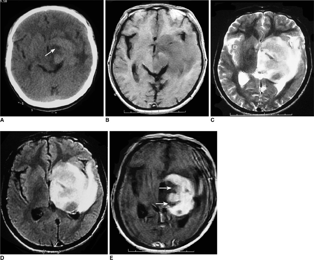
Fig. 1 Images of 52-year-old man with lymphoma of left basal ganglia including plain CT image (A), axial T1-weighted image (B), T2-weighted image (C), FLAIR (D) and postcontrast axial T1-weighted image (E). Abnormally deep depression at tumor margin (notch sign) (arrow) was found on post-contrast axial T1-weighted image and plain CT image.
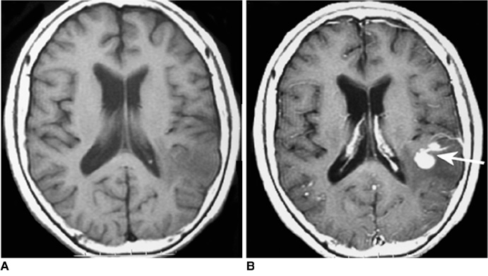
Fig. 2 Images of 71-year-old woman with lymphoma of left temporal lobe including pre-contrast axial T1-weighted image (A), post-contrast axial T1-weighted image (B). 'Notch sign' (arrow) was found on post-contrast T1-weighted image.
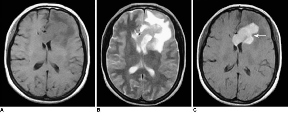
Fig. 3 Images of 49-year-old woman with primary central nervous system lymphoma including pre-contrast axial T1-weighted image (A), pre-contrast T2-weighted image (B), post-contrast axial T1-weighted image (C). 'Notch sign' (arrow) was found after contrast injection.
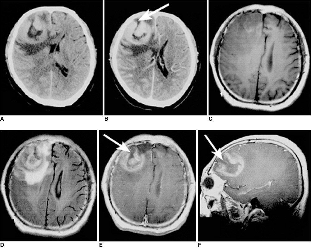
Fig. 4 Images of 57-year-old man with lymphoma of right frontal lobe including plain CT image (A), contrast CT image (B), axial T1-weighted image (C), FLAIR (D), postcontrast axial T1-weighted image (E) and sagittal T1-weighted image (F). On plain CT image; lesion was hyperdense. 'Open-ring' enhancement (arrows) was found and was characterized as thick and not uniform.

Fig. 5 Images of 56-year-old woman with lymphoma involving frontal lobe bilaterally including pre-contrast axial T1-weighted image (A), pre-contrast T2-weighted image (B) and post-contrast axial T1-weighted image (C). 'Open-ring' enhancement (arrow) was found and was characterized as thick and not uniform.
Reference
-
1. Gutmann J, Kendall B. Unusual appearances of primary central nervous system non-Hodgkin's lymphoma. Clin Radiol. 1994. 49:696–702.2. Watanabe M, Tanaka R, Takeda N, Wakabayashi K, Takahashi H. Correlation of computed tomography with the histopathology of primary malignant lymphoma of the brain. Neuroradiology. 1992. 34:36–42.3. Coulon A, Lafitte F, Hoang-Xuan K, Martin-Duverneuil N, Mokhtari K, Blustajn J, et al. Radiographic findings in 37 cases of primary CNS lymphoma in immunocompetent patients. Eur Radiol. 2002. 12:329–340.4. Miller DC, Hochberg FH, Harris NL, Gruber ML, Louis DN, Cohen H. Pathology with clinical correlations of primary central nervous system non-Hodgkin's lymphoma. The Massachusetts General Hospital experience 1958-1989. Cancer. 1994. 74:1383–1397.5. Nitta T, Uda K, Ebato M, Ikezaki K, Fukui M, Sato K. Primary peripheral-postthymic T-cell lymphoma in the central nervous system: immunological and molecular approaches to diagnosis. J Neurosurg. 1995. 82:77–82.6. Slone HW, Blake JJ, Shah R, Guttikonda S, Bourekas EC. CT and MRI findings of intracranial lymphoma. AJR Am J Roentgenol. 2005. 184:1679–1685.7. Jack CR Jr, Reese DF, Scheithauer BW. Radiographic findings in 32 cases of primary CNS lymphoma. AJR Am J Roentgenol. 1986. 146:271–276.8. Jack CR Jr, O'Neill BP, Banks PM, Reese DF. Central nervous system lymphoma: histologic types and CT appearance. Radiology. 1988. 167:211–215.9. Johnson BA, Fram EK, Johnson PC, Jacobowitz R. The variable MR appearance of primary lymphoma of the central nervous system: comparison with histopathologic features. AJNR Am J Neuroradiol. 1997. 18:563–572.10. Küker W, Nägele T, Korfel A, Heckl S, Thiel E, Bamberg M, et al. Primary central nervous system lymphomas (PCNSL): MRI features at presentation in 100 patients. J Neurooncol. 2005. 72:169–177.11. Jenkins CN, Colquhoun IR. Characterization of primary intracranial lymphoma by computed tomography: an analysis of 36 cases and a review of the literature with particular reference to calcification haemorrhage and cyst formation. Clin Radiol. 1998. 53:428–434.12. Onda K, Wakabayashi K, Tanaka R, Takahashi H. Intracranial malignant lymphomas: clinicopathological study of 26 autopsy cases. Brain Tumor Pathol. 1999. 16:29–35.13. Bataille B, Page P. Prognostic factors and primary cerebral lymphoma. Neurochirurgie. 1997. 43:385–387. [French].14. Dubuisson A, Kaschten B, Lénelle J, Martin D, Robe P, Fassotte MF, et al. Primary central nervous system lymphoma report of 32 cases and review of the literature. Clin Neurol Neurosurg. 2004. 107:55–63.15. Herrlinger U, Schabet M, Bitzer M, Petersen D, Krauseneck P. Primary central nervous system lymphoma: from clinical presentation to diagnosis. J Neurooncol. 1999. 43:219–226.16. Erdag N, Bhorade RM, Alberico RA, Yousuf N, Patel MR. Primary lymphoma of the central nervous system: typical and atypical CT and MR imaging appearances. AJR Am J Roentgenol. 2001. 176:1319–1326.17. Gliemroth J, Kehler U, Gaebel C, Arnold H, Missler U. Neuroradiological findings in primary cerebral lymphomas of non-AIDS patients. Clin Neurol Neurosurg. 2003. 105:78–86.18. Cellerier P, Chiras J, Gray F, Metzger J, Bories J. Computed tomography in primary lymphoma of the brain. Neuroradiology. 1984. 26:485–492.19. Hochberg FH, Miller DC. Primary central nervous system lymphoma. J Neurosurg. 1988. 68:835–853.20. Holodny AI, Nusbaum AO, Festa S, Pronin IN, Lee HJ, Kalnin AJ. Correlation between the degree of contrast enhancement and the volume of peritumoral edema in meningiomas and malignant gliomas. Neuroradiology. 1999. 41:820–825.21. Koeller KK, Smirniotopoulos JG, Jones RV. Primary central nervous system lymphoma: radiologic-pathologic correlation. Radiographics. 1997. 17:1497–1526.22. Brown JH, Stallmeyer MJ, Lustrin ES, Chew FS. Primary cerebral lymphoma. AJR Am J Roentgenol. 1995. 165:626.23. Reni M, Ferreri AJ, Garancini MP, Villa E. Therapeutic management of primary central nervous system lymphoma in immunocompetent patients: results of a critical review of the literature. Ann Oncol. 1997. 8:227–234.24. Schabet M. Epidemiology of primary CNS lymphoma. J Neurooncol. 1999. 43:199–201.25. Jiddane M, Nicoli F, Diaz P, Bergvall U, Vincentelli F, Hassoun J, et al. Intracranial malignant lymphoma. Report of 30 cases and review of the literature. J Neurosurg. 1986. 65:592–599.26. Thomas M, MacPherson P. Computed tomography of intracranial lymphoma. Clin Radiol. 1982. 33:331–336.27. DeAngelis LM. Cerebral lymphoma presenting as a nonenhancing lesion on computed tomographic/magnetic resonance scan. Ann Neurol. 1993. 33:308–311.28. Terae S, Ogata A. Nonenhancing primary central nervous system lymphoma. Neuroradiology. 1996. 38:34–37.29. Braks E, Urbach H, Pels H, Träber F, Block W, Schild HH. Primary central nervous system immunocytoma: MRI and spectroscopy. Neuroradiology. 2000. 42:738–741.30. Masdeu JC, Quinto C, Olivera C, Tenner M, Leslie D, Visintainer P. Open-ring imaging sign: highly specific for atypical brain demyelination. Neurology. 2000. 54:1427–1433.31. Bizzi A, Movsas B, Tedeschi G, Phillips CL, Okunieff P, Alger JR, et al. Response of non-Hodgkin lymphoma to radiation therapy: early and long-term assessment with H-1 MR spectroscopic imaging. Radiology. 1995. 194:271–276.
- Full Text Links
-
- Actions
-
Cited
- CITED
-
- Close
- Share
-
- Similar articles
-
- Correspondence Re: MRI Findings of Primary CNS Lymphoma in 26 Immunocompetent Patients
- CNS Involvement in the Non-odgkin's Lymphoma
- Primary Central Nervous System Lymphoma Associated with Systemic Lymphoma: Case Report
- Primary CNS Lymphoma in Immunocompetent Patients A Clinical and Pathological Study
- MRI findings of primary CNS lymphoma
