Bronchus-Associated Lymphoid Tissue (BALT) Lymphoma of the Lung Showing Mosaic Pattern of Inhomogeneous Attenuation on Thin-section CT: A Case Report
- Affiliations
-
- 1Department of Radiology, Hallym University Sacred Heart Hospital, Anyang City, Kyungki-do, Korea. ijlee@www.hallym.or.kr
- KMID: 877076
- DOI: http://doi.org/10.3348/kjr.2000.1.3.159
Abstract
- The authors present a case of histologically proven bronchus-associated lymphoid tissue (BALT) lymphoma of the lung in a patient with primary Sjogren's syn-drome that manifested on thin-section CT scan as a mosaic pattern of inhomoge-neous attenuation due to mixed small airway and infiltrative abnormalities
Keyword
MeSH Terms
Figure
-
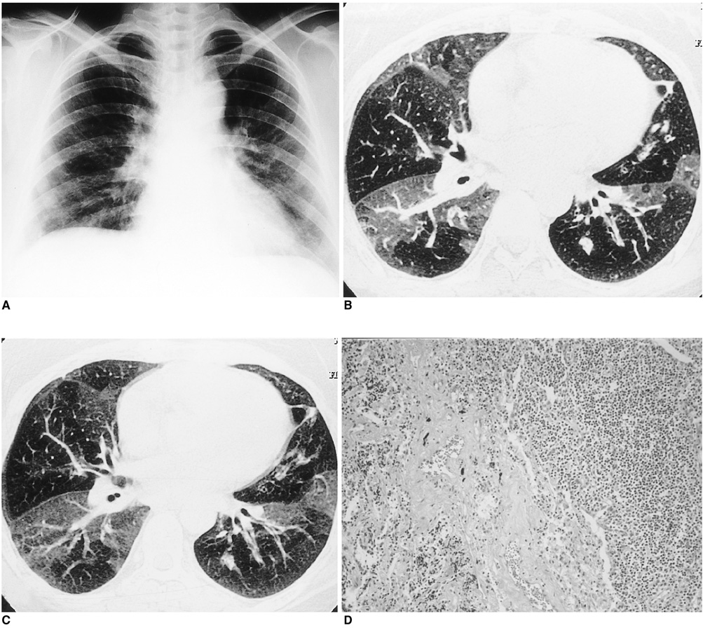
Fig. 1 BALT lymphoma of the lung in a 33-year-old housewife with primary Sjögren's syndrome. A. Posterior-anterior chest radiograph shows diffuse reticulonodular densities in both lungs. B. Inspiratory thin-section CT scan shows mosaic pattern of inhomogeneous attenuation. Note the smaller caliber of pulmonary vessels within the area of lower attenuation than within the higher. Note too the presence of bronchial wall thickening in the lingular division of the upper lobe of the left lung. C. Expiratory thin-section CT scan shows no difference in the extent of mosaic pattern of inhomogeneous attenuation. D. Light microscopic examination of resected specimen shows diffuse infiltration of marginal zone lymphocytes into peribronchiolar alveolar space (H and E, × 100).
Cited by 1 articles
-
Mucosa-Associated Lymphoid Tissue Lymphoma of the Esophagus Coexistent with Bronchus-Associated Lymphoid Tissue Lymphoma of the Lung
Jae-Joon Chung, Myeong-Jin Kim, Jeong-Hae Kie, Ki Whang Kim
Yonsei Med J. 2005;46(4):562-566. doi: 10.3349/ymj.2005.46.4.562.
Reference
-
1. Lee DK, Im J-G, Lee KS, et al. B-cell lymphoma of bronchus-associated lymphoid tissue (BALT): CT features in 10 patients. J Comput Assist Tomogr. 2000. 24:30–34.2. O'Donnell PG, Jackson SA, Tung KT, Hassan B, Wilkins B, Mead GM. Radiological appearance of lymphomas arising from mucosa-associated lymphoid tissue (MALT) in the lung. Clin Radiol. 1998. 53:258–263.3. Knisely BL, Mastey LA, Mergo PJ, et al. Pulmonary mucosa-associated lymphoid tissue lymphoma: CT and pathologic findings. AJR. 1999. 172:1321–1326.4. Wislez M, Cadranel J, Antoine M, et al. Lymphoma of pulmonary mucosa-associated lymphoid tissue: CT scan findings and pathological correlations. Eur Respir J. 1999. 14:423–429.5. McCulloch GL, Sinnatamby R, Stewart S, Goddard M, Flower CDR. High-resolution computed tomographic appearance of MALToma of the lung. Eur Radiol. 1998. 8:1669–1673.6. Chow WH, Ducheine Y, Hilfer J, Brandstetter RD. Chronic pneumonia. Primary malignant non-Hodgkin's lymphoma of the lung arising in mucosa-associated lymphoid tissue. Chest. 1996. 110:838–840.7. Franquet T, Gimenez A, Monill JM, Diaz C, Geli C. Primary Sjögren's syndrome and associated lung disease: CT findings in 50 patients. AJR. 1997. 169:655–658.8. Hansen LA, Parkash UBS, Colby TV. Pulmonary lymphoma in Sjögren's syndrome. Mayo Clin Proc. 1989. 64:920–931.9. Worthy SA, Müller NL, Hartman TE, Swensen SJ, Padley SPG, Hansell DM. Mosaic attenuation pattern on thin-section CT scans of the lung: differentiation among infiltrative lung, airway, and vascular disease as a cause. Radiology. 1997. 205:465–470.10. Shah RM, Sexauer W, Ostrum BJ, et al. High-resolution CT in the acute exacerbation of cystic fibrosis: evaluation of acute findings, reversibility of those findings, and clinical correlation. AJR Am J Roentgenol. 1997. 169:375–380.
- Full Text Links
-
- Actions
-
Cited
- CITED
-
- Close
- Share
-
- Similar articles
-
- A Case of Bronchus-Associated Lymphoid Tissue(BALT) Lymphoma in the Lung of the Patient with Primary Sjogren's Syndrome
- Cases of the Pulmonary Malignant Lymphoma of the Bronchus-Associated Lymphoid Tissue (BALT)
- Mucosa-Associated Lymphoid Tissue Lymphoma of the Esophagus Coexistent with Bronchus-Associated Lymphoid Tissue Lymphoma of the Lung
- A case of bronchus-associated lymphoid tissue (BALT) lymphoma in the patient with rheumatoid arthritis
- Radiation Therapy for Bronchial Associated Lymphoid Tissue (BALT) Lymphoma : A case report
