Evaluation of Tracheobronchial Diseases: Comparison of Different Imaging Techniques
- Affiliations
-
- 1Department of Radiology, Seoul National University College of Medicine and the Institute of Radiation Medicine, SNUMRC, Seoul, Korea. imjg@radcom.snu.ac.kr
- KMID: 877073
- DOI: http://doi.org/10.3348/kjr.2000.1.3.135
Abstract
OBJECTIVE
To compare the clinical utility of the different imaging techniques used for the evaluation of tracheobronchial diseases. MATERIALS AND METHODS: Forty-one patients with tracheobronchial diseases [tuberculosis (n = 18), bronchogenic carcinoma (n = 10), congenital abnormality (n = 3), post-operative stenosis (n = 2), and others (n = 8)] underwent chest radi-ography and spiral CT. Two sets of scan data were obtained: one from routine thick-section axial images and the other from thin-section axial images. Multiplanar reconstruction (MPR) and shaded surface display (SSD) images were obtained from thin-section data. Applying a 5-point scale, two observers compared chest radiography, routine CT, thin-section spiral CT, MPR and SSD imaging with regard to the detection, localization, extent, and characterization of a lesion, information on its relationship with adjacent structures, and overall information. RESULTS: SSD images were the most informative with regard to the detection (3.95 +/-0.31), localization (3.95 +/-0.22) and extent of a lesion (3.85 +/-0.42), and overall information (3.83 +/-0.44), while thin-section spiral CT scans provided most information regarding its relationship with adjacent structures (3.56 +/-0.50) and characterization of the lesion (3.51 +/-0.61). CONCLUSION: SSD images and thin-section spiral CT scans can provide valuable information for the evaluation of tracheobronchial disease.
Keyword
MeSH Terms
Figure
-

Fig. 1 Bronchogenic carcinoma in a 66-year-old man. A. Coronal MPR image shows apparent total obstruction of the left main bronchus by a polypoid soft tissue lesion (arrow). B. Curved reformatted image along the axis of the left upper lobe shows focal wall thickening of the left main bronchus (arrow).
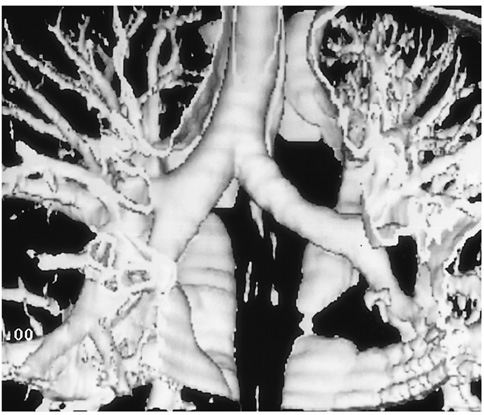
Fig. 2 SSD image from which unnecessary lung was removed shows the trachea and bilateral main bronchi.

Fig. 3 SSD image preprocessed with slice by slice editing to remove unwanted structures shows complete focal obstruction of the left upper lobar bronchus (arrow).
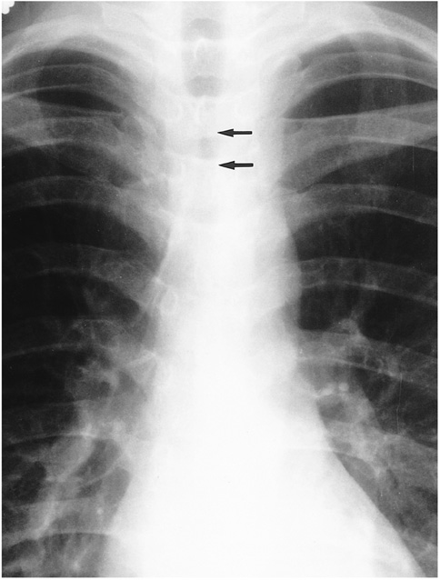
Fig. 4 Endotracheal tuberculosis in a 26-year-old woman. Posteroanterior chest radiograph shows segmental narrowing of the trachea (arrows).

Fig. 5 Endobronchial tuberculosis in a 67-year-old woman. A. Thin-section spiral CT shows mild irregular wall thickening (arrow) of the right main bronchus (score for detection = 4). B. SSD image shows subtle irregularity (arrow) in the right main bronchus (score for detection = 2).
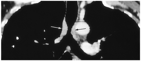
Fig. 6 Tracheal tumor in a 54-year-old man. The histology of this tumor was not confirmed because its hypervascularity, as revealed by bronchoscopy, precluded biopsy. Coronal MPR image demonstrates the extent of concentric wall thickening of the trachea (arrows).
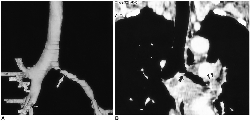
Fig. 7 Endobronchial tuberculosis in a 55-year-old woman. SSD image (A) (score for extent = 4) and coronal MPR image (B) (score for extent =2) show irregular narrowing of the left main bronchus (arrow) and complete obstruction of the left upper lobar bronchus (arrowheads).

Fig. 8 An outpouching lesion at the anastomotic site after sleeve left upper lobectomy in a 67-year-old man. SSD (A), routine enhanced CT (B), and coronal MPR image (C) show an outpouching lesion at the anastomotic site (arrow). For these three techniques, the respective score for relationship was 0, 4, and 3, respectively.
Cited by 1 articles
-
Usefulness of Digital Tomosynthesis for the Detection of Airway Obstruction: A Case Report of Bronchial Carcinosarcoma
Sung-Joon Park, Ji Yung Choo, Ki Yeol Lee, Je-Hyeong Kim, Jung-Woo Choi, Suk Keu Yeom, Baek Hyun Kim
Cancer Res Treat. 2015;47(3):544-548. doi: 10.4143/crt.2013.220.
Reference
-
1. Quint LE, Whyte RI, Kazerooni EA, et al. Stenosis of the central airways: evaluation by using helical CT with multiplanar reconstruction. Radiology. 1995. 194:871–877.2. Sagy M, Poustchi-Amin M, Nimkoff L, Silver P, Shikowitz M, Leonidas JC. Spiral computed tomographic scanning of the chest with three dimensional imaging in the diagnosis and management of paediatric intrathoracic airway obstruction. Thorax. 1996. 51:1005–1009.3. LoCicero J, Costello P, Campos CT, et al. Spiral CT with multiplanar and three-dimensional reconstructions accurately predicts tracheobronchial pathology. Ann Thorac Surg. 1996. 62:811–817.4. Kauczor HU, Wolcke B, Fischer B, Mildenberger P, Lorenz J, Thelen M. Three-dimensional helical CT of the tracheobronchial tree: evaluation of imaging protocols and assessment of suspected stenoses with bronchoscopic correlation. AJR. 1996. 167:419–424.5. Lacrosse M, Trigaux JP, Van Beers BE, Weynants P. 3D spiral CT of the tracheobronchial tree. J Comput Assist Tomogr. 1995. 19:341–347.6. Gurney JW. Missed lung cancer at CT: imaging findings in nine patients. Radiology. 1996. 199:117–122.7. Whyte RI, Quint LE, Kazerooni EA, Cascade PN, Iannettoni MD, Orringer MB. Helical computed tomography for the evaluation of tracheal stenosis. Ann Thorac Surg. 1995. 60:27–31.8. Hernandez RJ, Tucker GF. Congenital tracheal stenoses: role of CT and high kV films. Pediatr Radiol. 1987. 17:192–196.9. Lee KS, Yoon JH, Kim TK, Kim JS, Chung MP, Kwon OJ. Evaluation of tracheobronchial disease with helical CT with multiplanar and three-dimensional reconstruction: correlation with bronchoscopy. RadioGraphics. 1997. 17:555–567.10. Naidich DP, Gruden JF, McGuinness G, McCauley DI, Bhalla M. Volumetric (helical/spiral) CT (VCT) of the airways. J Thorac Imag. 1998. 12:11–28.11. Boiselle PM, Patz EF Jr, Vining DJ, Weissleder R, Shepard JA, McLoud TC. Imaging of mediastinal lymph nodes: CT, MR, and FDG PET. RadioGraphics. 1998. 18:1061–1069.12. Higgins WE, Ramaswamy K, Swift RD, McLennan G, Hoffman EA. Virtual bronchoscopy for three-dimensional pulmonary image assessment: state of the art and future needs. RadioGraphics. 1998. 18:761–778.
- Full Text Links
-
- Actions
-
Cited
- CITED
-
- Close
- Share
-
- Similar articles
-
- Effect of Anesthetics on Protein Content of Alveolar Washings of Rabbits
- Advanced Imaging Techniques for Assessing Fat, Iron, and Fibrosis in Chronic Liver Disease
- Comparison of Cine Magnetic Resonance Imaging with Doppler Echocardiography for the Quantative Evaluation of Tricuspid Regurgitation in Newborn
- Recent development of diagnostic imaging of hepatocellular carcinoma
- Advances in the Endoscopic Assessment of Inflammatory Bowel Diseases: Cooperation between Endoscopic and Pathologic Evaluations
