Treatment of Branch Retinal Artery Occlusion With Transluminal Nd:YAG Laser Embolysis
- Affiliations
-
- 1Kong's Eye Clininc, Seoul, Korea.
- 2Department of Ophthalmology, Asan Medical Center, University of Ulsan College of Medicine, Seoul, Korea. Junekim@amc.seoul.kr
- KMID: 754772
- DOI: http://doi.org/10.3341/kjo.2009.23.4.315
Abstract
- The purpose of this paper was to report a successful treatment of transluminal Nd:YAG laser embolysis (NYE) for branch retinal artery occlusion (BRAO) with visible emboli. Two patients with acute, severe vision loss secondary to a branch retinal artery occlusion with visible emboli in one eye underwent NYE. A complete ocular examination was performed which included biomicroscopy of the posterior pole of the retina, intraocular pressure measurement, fundus color photographs, and fluorescein angiography (FA). After the NYE, the two patients showed dramatic improvements in best-corrected visual acuity, as well as, immediate and dramatic restorations in flow past the obstructed arteriole in FA. NYE is a treatment modality to be considered in patients with BRAO who present acutely with severe vision loss and a visible embolus.
MeSH Terms
Figure
-
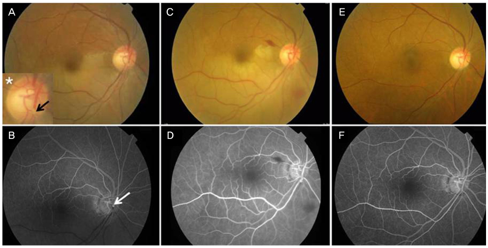
Fig. 1 Color fundus photographs and fluorescein angiography (FA) showing branch retinal artery obstruction in case 1. (A) Color fundus photograph demonstrating an intraluminal embolus (arrow) and whitening of the retina along the inferior and temporal vascular arcades. inserted: the asterisk panel '*' shows a magnification of the embolus. (B) FA of the same patient showing obstructed blood flow at the site of the embolus and a downstream filling defect. (C) Color fundus photograph of the same patient taken five days after Nd:YAG laser embolysis (NYE). Note the small amount of vitreous hemorrhage around the disc. (D) Fluorescein angiography (FA) of the same patient five days after NYE showing perfusion at the site of the embolus and restoration of the downstream blood flow. (E) Color fundus photograph taken three months after NYE. There was no remaining vitreous hemorrhage. (F) FA at three months after NYE showing a patent retinal artery with good blood flow and no neovascularization.
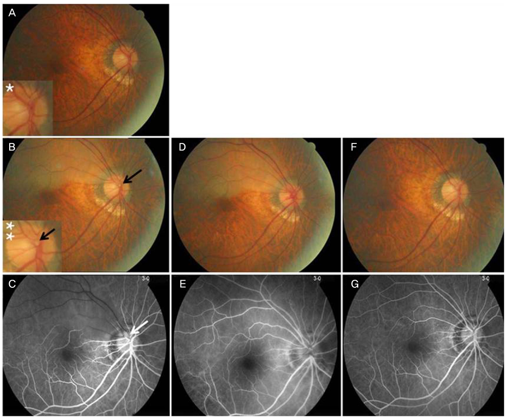
Fig. 2 Color fundus photographs and fluorescein angiography showing branch retinal artery obstruction (BRAO) in Case 2. (A) Color fundus photograph six mon prior to BRAO. The asterisk panel '*' shows a magnification of the disc and indicates no transluminal embolus. (B) Color fundus photograph showing a superior temporal branch retinal artery occlusion. Note the transluminal embolus (arrow) and opacification of the retina along the superior and temporal vascular arcades. The double asterisk panel '**' shows a magnification of the transluminal embolus. (C) Fluorescein angiography (FA) showing the filling defect in the arteriole corresponding to the intraluminal embolus. (D) Color fundus photograph taken three days after Nd:YAG laser embolysis (NYE). Note the small amount of vitreous hemorrhage around the disc. (E) FA three days after NYE showing restoration of retinal blood flow. (F) Color fundus photograph taken two mon after NYE. Note the spontaneous clearing of the small vitreous hemorrhage. (G) FA two mon after NYE showing a return of retinal blood flow.
Cited by 2 articles
-
Treatment of Acute Central Retinal Artery Occlusion with Ocular Ischemic Syndrome
Jong Hwan Lee, Ho Seok Moon, Dong Heun Nam, Dae Yeong Lee
J Korean Ophthalmol Soc. 2014;55(8):1242-1247. doi: 10.3341/jkos.2014.55.8.1242.A Case of Idiopathic Pediatric Acute Branch Retinal Artery Occlusion Involving the Macular Area
Ji Hye Jang, Jong Won Moon, Young Wook Cho
J Korean Ophthalmol Soc. 2014;55(2):304-308. doi: 10.3341/jkos.2014.55.2.304.
Reference
-
1. Brown GC, Shields JA. Cilioretinal arteries and retinal arterial occlusion. Arch Ophthalmol. 1979. 97:84–92.2. Ros MA, Larry ME, Uram M. Branch retinal-artery obstruction: a review of 201 eyes. Ann Ophthalmol. 1989. 21:103–107.3. Opremcak EM, Benner JD. Translumenal Nd:YAG laser embolysis for branch retinal artery occlusion. Retina. 2002. 22:213–216.4. Mason JO 3rd, Nixon PA, Albert MA Jr. Trans-luminal Nd:YAG laser embolysis for branch retinal artery occlusion. Retina. 2007. 27:573–577.5. Mason JO 3rd, Shah AA, Vail RS, et al. Branch retinal artery occlusion: visual prognosis. Am J Ophthalmol. 2008. 146:455–457.
- Full Text Links
-
- Actions
-
Cited
- CITED
-
- Close
- Share
-
- Similar articles
-
- An Electroretinographic Changes of Laser-induced Experimental Branch Retinal Vein Occlusion in the Rabbits
- Central Retinal Artery Occlusion Masquerading as Branch Retinal Artery Occlusion
- A Clinical Study of 36 Cases of Retinal Artery Occlusion
- Treatment of Acute Central Retinal Artery Occlusion with Ocular Ischemic Syndrome
- Retinal Arteriolar Changes in a Patient with Branch Retinal Vein Occlusion
