Korean J Ophthalmol.
2016 Oct;30(5):390-391. 10.3341/kjo.2016.30.5.390.
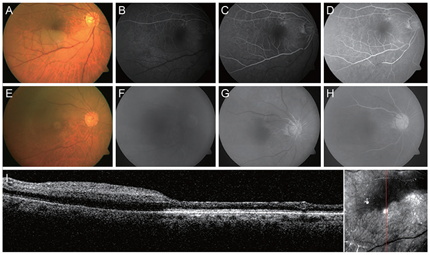
Central Retinal Artery Occlusion Masquerading as Branch Retinal Artery Occlusion
- Affiliations
-
- 1Department of Ophthalmology, Dongsan Medical Center, Keimyung University School of Medicine, Daegu, Korea. eyedr@dsmc.or.kr
- KMID: 2353833
- DOI: http://doi.org/10.3341/kjo.2016.30.5.390
Abstract
- No abstract available.
MeSH Terms
Figure
Reference
-
1. Hayreh SS, Zimmerman MB. Fundus changes in central retinal artery occlusion. Retina. 2007; 27:276–289.2. Ahn SJ, Woo SJ, Park KH, et al. Retinal and choroidal changes and visual outcome in central retinal artery occlusion: an optical coherence tomography study. Am J Ophthalmol. 2015; 159:667–676.3. Hayreh SS. Ocular vascular occlusive disorders: natural history of visual outcome. Prog Retin Eye Res. 2014; 41:1–25.4. Chen SN, Hwang JF, Chen YT. Macular thickness measurements in central retinal artery occlusion by optical coherence tomography. Retina. 2011; 31:730–737.
- Full Text Links
- Actions
-
Cited
- CITED
-
- Close
- Share
- Similar articles
-
- A Clinical Study of 36 Cases of Retinal Artery Occlusion
- Incomplete Central Retinal Artery Occlusion
- A Case of Cilioretinal Artery Occlusion Associated with Central Retinal Vein Occlusion
- The Successful Treatment of a Case of Central Retinal Artery Occlusion
- Central Retinal Artery Occlusion after Cervical Spine Surgery in Prone Position: A Case Report


