Infantile Vitreous Hemorrhage as the Initial Presentation of X-linked Juvenile Retinoschisis
- Affiliations
-
- 1Department of Ophthalmology, Seoul National University College of Medicine, Seoul, Korea. ysyu@snu.ac.kr
- 2Department of Laboratory Medicine, Seoul National University Hospital, Seoul, Korea.
- 3Seoul Artificial Eye Center, Clinical Research Institute, Seoul National University Hospital, Seoul, Korea.
- KMID: 754730
- DOI: http://doi.org/10.3341/kjo.2009.23.2.118
Abstract
- The authors report two cases of X-linked juvenile retinoschisis (XLRS) manifested as bilateral vitreous hemorrhage as early as in an 1-month-old infant and in a 3-month-old infant. The one-month-old male infant showed massive bilateral vitreous hemorrhage. During vitrectomy, thin membrane representing an inner part of schisis cavity was excised and intraschisis hemorrhage was evacuated. As intraschisis cavities were cleared, the stump of inner layer appeared as the demarcation line between the outer layer of the schisis retina and non-schisis retina. The other three-month-old male infant presenting with esodeviation also showed bilateral vitreous hemorrhage. Typical bilateral retinoschisis involving maculae could be seen through vitreous hemorrhage in both eyes on fundus examination. Spontaneous absorption of hemorrhage was observed on regular follow-up. XLRS could be manifested as massive hemorrhage inside or outside of the schisis cavity early in infancy.
MeSH Terms
Figure
-
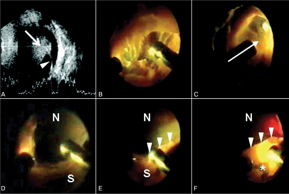
Fig. 1 Preoperative ultrasonography (USG) and intraoperative fundus findings of the right eye in case 1. USG shows vitreous hemorrhage (arrow) and suggests retinoschisis (arrowhead) (A). Massive vitreous hemorrhage is being removed after lensectomy (B). Thin membranous structure (arrow) is detected during careful vitrectomy (C). After excision of thin membranous inner layer, nonschisis retina (N) can be shown (D). The stump (arrowheads) of inner layer can be shown with adherent intraschisis blood clot (E). As intraschisis blood is being removed, the outer layer (star) of the schisis retina (S) appears (F).

Fig. 2 Fundus findings observed at the age of 32 months in case 2. Bilateral retinoschisis involving posterior pole can be shown (A: right eye, B: left eye). Fundus findings were stationary without progression with partially remnant hemorrhage in the right eye at the age of 32 months.
Reference
-
1. Prasad A, Wagner R, Bhagat N. Vitreous hemorrhage as the initial manifestation of X-linked retinoschisis in a 9-month-old infant. J Pediatr Ophthalmol Strabismus. 2006. 43:56–58.2. George ND, Yates JR, Bradshaw K, Moore AT. Infantile presentation of X linked retinoschisis. Br J Ophthalmol. 1995. 79:653–657.3. Lee JJ, Kim JH, Choung HK, et al. Long-term results of vision and fundus findings in X-linked juvenile retinoschisis. J Korean Ophthalmol Soc. 2007. 48:911–918.4. Ortiz JM, Yanoff M, Cameron D, Schaffer D. Disseminated intravascular coagulation in infancy and in the neonate. Arch Ophthalmol. 1982. 100:1413–1415.5. Functional implications of the spectrum of mutations found in 234 cases with X-linked juvenile retinoschisis. Hum Mol Genet. 1998. 7:1185–1192.6. Kaur B, Taylor D. Fundus hemorrhages in infancy. Surv Ophthalmol. 1992. 37:1–17.
- Full Text Links
-
- Actions
-
Cited
- CITED
-
- Close
- Share
-
- Similar articles
-
- Bilateral Juvenile Retinoschisis in four Brothers of a Family
- Long-term Results of Vision and Fundus Findings in X-linked Juvenile Retinoschisis
- A novel mutation in XLRS1 gene in X-linked juvenile retinoschisis
- A Novel Mutation in the XLRS1 Gene in a Korean Family with X-linked Retinoschisis
- Differential Diagnosis in the Rhegmatogenous Retinal Detachment
