A Case of Radiation Retinopathy of Left Eye After Radiation Therapy of Right Brain Metastasis
- Affiliations
-
- 1Department of Ophthalmology, College of Medicine, Dongsan Medical Center, Keimyung University, Daegu, Korea. changsd@dsmc.or.kr
- KMID: 754729
- DOI: http://doi.org/10.3341/kjo.2009.23.2.114
Abstract
- A 37-year-old female, who had received modified radical mastectomy for cancer of her right breast, presented with decreased visual acuity in the left eye after radiation therapy for the management of the metastasis to her right brain 14 months ago. After ocular examination, we diagnosed her as radiation retinopathy. At the time of the first visit, the corrected best visual acuity was 0.4 in the left eye, and fundus examination revealed cotton wool spots and cystoid macular edema (CME). The findings in the right eye were normal except for cotton wool spots in the superior major arch. Fluorescein angiography (FA) showed marked telangiectasia and microaneurysms in her left eye but tiny microaneurysms in her right eye. Subsequent optical coherent tomography (OCT) showed CME. We injected intravitreal triamcinolone acetonide (TA). Two weeks after treatment, the visual acuity was improved to 0.6 and the retinal thickness was decreased. Three months later, the visual acuity in the left eye was dropped to 0.3 due to the recurrence of CME, so we injected intravitreal TA again. Five months later, visual acuity was improved to 0.5 and OCT revealed the improvement of CME. The incidence of radiation retinopathy is higher in the side nearer to radiation, but careful radiation blocking is also required on the opposite side of irradiation site considering the possibility of radiation retinopathy and careful observation is required on both sides of the eyes when performing fundus examination.
MeSH Terms
-
Adult
Brain Neoplasms/*radiotherapy/secondary
Breast Neoplasms/pathology/radiotherapy/surgery
Diagnosis, Differential
Female
Fluorescein Angiography
Follow-Up Studies
Fundus Oculi
Glucocorticoids/administration & dosage
Humans
Radiation Injuries/diagnosis/drug therapy/*etiology
Retina/pathology/*radiation effects
Retinal Diseases/diagnosis/drug therapy/*etiology
Tomography, Optical Coherence
Triamcinolone Acetonide/administration & dosage
Figure
-
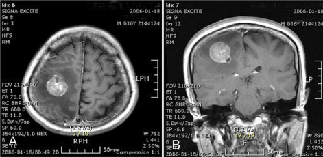
Fig. 1 Axial section (A) coronal section (B) of T1 weighted MRI shows the brain metastasis at the right frontal lobe.

Fig. 2 Color fundus photograph and fluorescein angiography of both eyes. (A) Color fundus photograph of the left eye at initial examination shows diffuse macular edema, cotton wool spots and tortuous retinal vessel at the superotemporal arcade. (B) Fluorescein angiography of left eye at initial examination shows vascular telangiectasis, and aneurysmal dilation of retinal vessels with leakage of dye. (C) Color fundus photograph of the right eye at initial examination shows suspected cotton wool spot near superotemporal arcade and other findings are unremarkable. (D) Fluorescein angiography of right eye at initial examination shows minimal microaneurysms without macular edema.
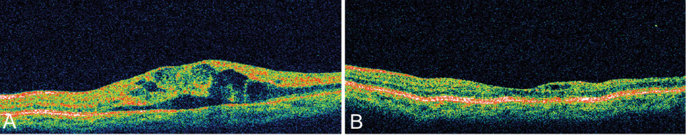
Fig. 3 (A) Optical coherence tomogram shows marked cystoid macula edema. (B) Five months after intravitreal triamcinolone acetonide injection, optical coherence tomogram shows decreased macular thickness and marked improvement of cystoid macula edema.
Reference
-
1. Cibis PA, Noell WK, Eichel B. Ocular effects produced by high intensity x-radiation. AMA Arch Ophtahlmol. 1955. 53:651–663.2. Boozalis GT, Schachat AP, Green WR. Subretinal neovascularization from the retina in radiation retinopathy. Retina. 1987. 7:156–161.3. Ching SV, Gillette SM, Powers BE, et al. Radiation-induced ocular injury in the dog: a histological study. Int J Radiat Oncol Biol Phys. 1990. 19:321–328.4. Chee PH. Radiation retinopathy. Am J Ophthalmol. 1968. 66:860–865.5. Brown GC, Shields JA, Sanborn G, et al. Radiation retinopathy. Ophthalmology. 1982. 89:1494–1501.6. Amoaku WM, Archer DB. Cephalic radiation and retinal vasculopathy. Eye. 1990. 4:195–203.7. Chan RC, Shukovsky LJ. Effects of irradiation on the eye. Radiology. 1976. 120:673–675.8. Stallard HB. Radiant energy as a pathogenic and a therapeutic agent in ophtahlmic disorder. Br J Ophthalmol. 1933. 6:1–126.9. Seong GJ, Kim HB, Kim SH. A case of radiation retinopathy. J Korean Ophthalmol Soc. 1987. 28:691–694.10. Merriam GR, Szechter A, Focht EF. The effects of ionizing radiations on the eye. Front Radiant Ther Oncol. 1972. 6:346–385.11. Letschert JGJ, Gonzalez D, Oskam J, et al. Results of radiotherapy in patients with Stage I orbital non-Hodgkins lymphoma. Radiother Onchol. 1991. 22:36–44.12. Parsons JT, Bova FJ, Fitzgerald CR, et al. Radiation retinopathy after external-beam irradiation: analysis of time-dose factors. Int J Radiation Oncology Biol Phys. 1994. 30:765–773.13. Perrers-Taylor M, Brinkley D, Reynold T. Chorioretinal damage as a complication of radiotheraphy. Acta Radiol Ther Phys Biol. 1965. 3:431–440.14. Nakissa N, Rubin P, Strohl R, Keys H. Ocular and orbital complication following radiation therapy of paranasal sinus malignancies and review of literature. Cancer. 1992. 51:980–986.15. Peterson IA, Kriss JP, McDougall IR, Donaldson SS. Prognostic factors in the radiotherapy of Graves' ophthalmopathy. Int J Radiat Oncol Biol Phys. 1990. 19:259–264.16. Bessell EM, Henk JM, Whitelocke RAF, Wright JE. Ocular morbidity after radiotherapy of orbital and conjunctival lymphoma. Eye. 1987. 1:90–96.17. Gass JDM. A fluorescein angiographic study of macular dysfunction secondary to retinal artery occlusion, collagen vascular disease, and vitritis. Arch Ophthalmol. 1968. 80:606–617.18. Chaudhuri PR, Austin DJ, Rosenthal AR. Treatment of radiation retinopathy. Br J Ophthalmol. 1981. 65:623–625.19. Kinyoun JL, Chittum ME, Wells CG. Photocoagulation treatment of radiation retinopathy. Am J Ophthalmol. 1988. 105:470–478.20. Kinyoun JL, Lawrence BS, Barlow WE. Proliferative radiation retinopathy. Arch Ophthalmol. 1996. 114:1097–1100.21. Martidis A, Duker JS, Greenberg PB, et al. Intravitreal triamcinolone for refractory diabetic macular edema. Ophthalmology. 2002. 109:920–927.22. Jonas JB, Hayler JK, Panda-Jonas S. Intravitreal injection of crystalline cortisone as adjunctive treatment of proliferative vitreoretinopathy. Br J Ophthalmol. 2000. 84:1064–1067.23. Park CH, Jaffe GJ, Fekrat S. Intravitreal triamcinolone acetonide in eyes with cystoid macular edema associated with central retinal vein occlusion. Am J Ophthalmol. 2003. 136:419–425.
