Korean J Ophthalmol.
2009 Jun;23(2):74-79. 10.3341/kjo.2009.23.2.74.
Edema of the Photoreceptor Layer in Vogt-Koyanagi-Harada Disease Observed Using High-Resolution Optical Coherence Tomography
- Affiliations
-
- 1Department of Ophthalmology, School of Medicine, Pusan National University, Busan, Korea. bsoum@pusan.ac.kr
- 2Department of Ophthalmology, College of Medicine, Inje University, Busan, Korea.
- KMID: 754721
- DOI: http://doi.org/10.3341/kjo.2009.23.2.74
Abstract
-
PURPOSE: To evaluate the characteristics of fluid accumulation in the uveitic stage of Vogt-Koyanagi-Harada (VKH) disease using high resolution optical coherence tomography (OCT3).
METHODS
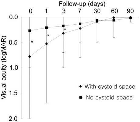
Twenty-eight eyes in 14 patients with VKH disease were reviewed retrospectively. These 28 eyes were divided into 19 eyes with intraretinal fluid (C group) and 9 eyes without intraretinal fluid (N group). Changes in visual acuity and fluid accumulation observed using OCT were compared between the two groups.
RESULTS
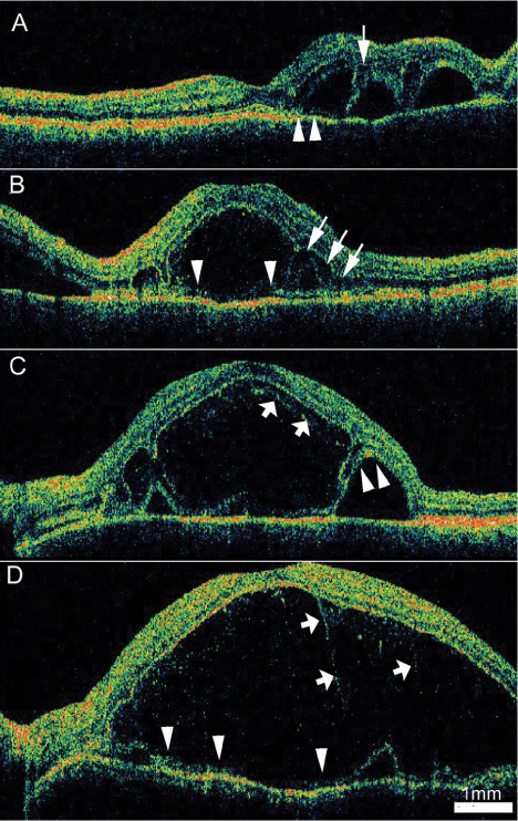
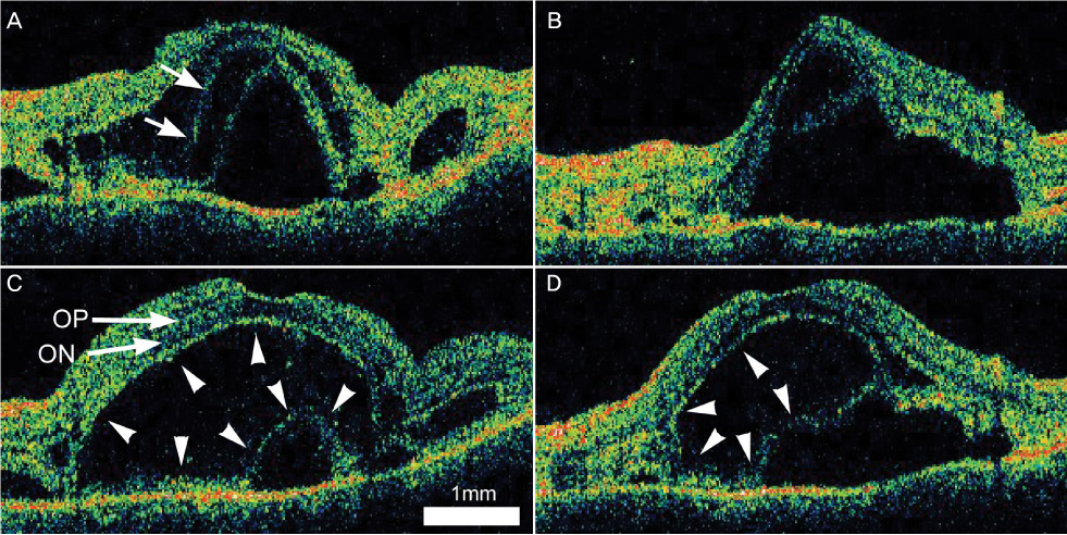
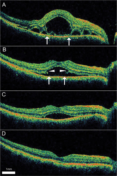
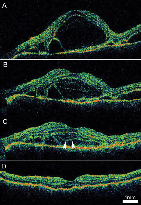
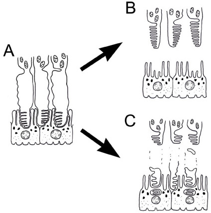
Visual acuity at the time of presentation was significantly worse in the C group than in the N group (p=0.005). The photoreceptor layer appeared to be double-layered due to a cystoid space in the C group. Layered structures and strands found in the cystoid space. Expanding sponge-form edema led to the development of a cystoid space in the photoreceptor layer. Intraretinal fluid resolved earlier than subretinal fluid. There were no observed differences in visual acuity between the two groups after four days of treatment.
CONCLUSIONS
Accumulation of intraretinal fluid was related to poor initial visual acuity, but not to final visual acuity. High resolution OCT findings indicate that edema of the photoreceptor layer participates in the development of a cystoid space.
Keyword
MeSH Terms
-
Adolescent
Adult
Diagnosis, Differential
Female
Follow-Up Studies
Humans
*Image Enhancement
Macular Edema/etiology/*pathology
Male
Middle Aged
Photoreceptor Cells, Vertebrate/*pathology
Retrospective Studies
Tomography, Optical Coherence/*methods
Uveomeningoencephalitic Syndrome/*complications/pathology
Young Adult
Figure
Cited by 1 articles
-
Clinical Features of Recurred Vogt-Koyanagi-Harada Syndrome during Oral Steroids Tapering Therapy
Ji Soo Kim, Dong Yoon Kim, Kyung Tae Kim, Ju Byung Chae
J Korean Ophthalmol Soc. 2019;60(4):331-339. doi: 10.3341/jkos.2019.60.4.331.
Reference
-
1. Read RW, Holland GN, Rao NA, et al. Revised diagnostic criteria for Vogt-Koyanagi-Harada disease: report of an international committee on nomenclature. Am J Ophthalmol. 2001. 131:647–652.2. Rubsamen PE, Gass JD. Vogt-Koyanagi-Harada syndrome. Clinical course, therapy, and long-term visual outcome. Arch Ophthalmol. 1991. 109:682–687.3. Parc C, Guenoun JM, Dhote R, Brezin A. Optical coherence tomography in the acute and chronic phases of Vogt-Koyanagi-Harada disease. Ocul Immunol Inflamm. 2005. 13:225–227.4. Maruyama Y, Kishi S. Tomographic features of serous retinal detachment in Vogt-Koyanagi-Harada syndrome. Ophthalmic Surg Lasers Imaging. 2004. 35:239–242.5. Hassenstein A, Bialasiewicz AA, Richard G. Optical coherence tomography in uveitis patients. Am J Ophthalmol. 2000. 130:669–670.6. Tsujikawa A, Yamashiro K, Yamamoto K, et al. Retinal cystoid spaces in acute Vogt-Koyanagi-Harada syndrome. Am J Ophthalmol. 2005. 139:670–677.7. de Smet MD, Rao NA. Retinal cystoid spaces in acute Vogt-Koyanagi-Harada syndrome. Am J Ophthalmol. 2005. 140:962–963.8. Yamaguchi Y, Otani T, Kishi S. Tomographic Features of Serous Retinal Detachment With Multilobular Dye Pooling in Acute Vogt-Koyanagi-Harada Disease. Am J Ophthalmol. 2007. 144:260–265.9. Ko TH, Fujimoto JG, Schuman JS, et al. Comparison of ultrahigh- and standard-resolution optical coherence tomography for imaging macular pathology. Ophthalmology. 2005. 112:1922.e1–1922.e15.10. Okamoto Y, Miyake Y, Horio N, et al. Delayed regeneration of foveal cone photopigments in Vogt-Koyanagi-Harada disease at the convalescent stage. Invest Ophthalmol Vis Sci. 2004. 45:318–322.11. Liem AT, Keunen JE, van Meel GJ, van Norren D. Serial foveal densitometry and visual function after retinal detachment surgery with macular involvement. Ophthalmology. 1994. 101:1945–1952.
- Full Text Links
- Actions
-
Cited
- CITED
-
- Close
- Share
- Similar articles
-
- Spectral-Domain Optical Coherence Tomography Findings of Vogt-Koyanagi-Harada Disease
- A Case of Vogt-Koyanagi-Harada-like Syndrome Associated with Immunotherapy (Pembrolizumab)
- Vogt-Koyanagi-Harada Syndrome Associated with Psoriasis Vulgaris
- A Case of Intravitreal Dexamethasone Implantation in a Patient with Vogt-Koyanagi-Harada Disease
- Acute Lymphoblastic Leukemia Manifesting as Acute Vogt-Koyanagi-Harada Disease

