Korean J Ophthalmol.
2009 Dec;23(4):325-328. 10.3341/kjo.2009.23.4.325.
Acute Lymphoblastic Leukemia Manifesting as Acute Vogt-Koyanagi-Harada Disease
- Affiliations
-
- 1Department of Ophthalmology, Seoul National University College of Medicine, Seoul Artificial Eye Center, Seoul National University Hospital Clinical Research Institute, Seoul, Korea. hgonyu@snu.ac.kr
- KMID: 754775
- DOI: http://doi.org/10.3341/kjo.2009.23.4.325
Abstract
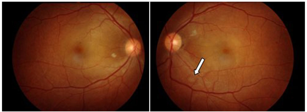
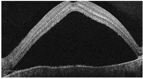
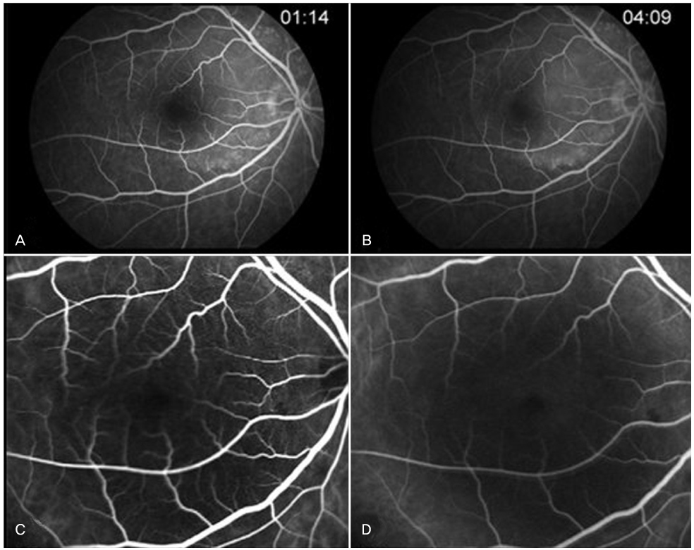
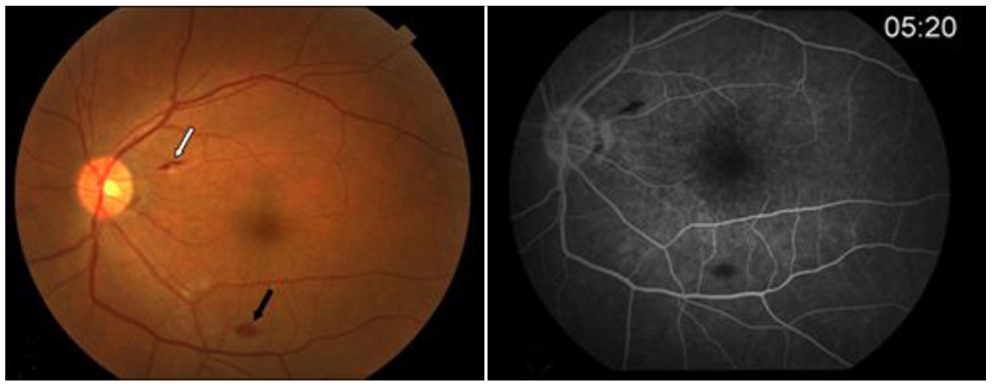
- We describe a case of bilateral exudative retinal detachment associated with prodromal symptoms simulating the presentation of acute Vogt-Koyanagi-Harada disease that was eventually diagnosed as acute lymphoblastic leukemia. A 42-year-old man presented with sudden visual loss in both eyes for two weeks. He complained of intermittent headache, neck stiffness and tinnitus for a month. His best-corrected visual acuities were 20/200 in both eyes. Fluorescein angiography, optical coherence topography and indocyanine green angiography featured bilateral serous retinal detachments. A clinical diagnosis of incomplete type Vogt-Koyanagi-Harada disease was considered. However, complete blood cell count showed a marked increase in the number of white blood cells and bone marrow examination revealed precursor B cell lymphoblastic leukemia. The patient started on induction chemotherapy. A week later, his best-corrected visual acuities were 20/25 and the serous retinal detachments were nearly absorbed in both eyes. Bilateral exudative retinal detachment associated with neurologic and auditory abnormalities may be a presenting sign of acute lymphoblastic leukemia. Clinicians should be aware of the possibility of leukemia in such patients.
MeSH Terms
Figure
Reference
-
1. Zimmerman LE, Thoreson HT. Sudden loss of vision in acute leukemia: a clinicopathologic report of two unusual cases. Surv Ophthalmol. 1964. 146:467–473.2. Stewart MW, Gitter KA, Cohen G. Acute leukemia presenting as a unilateral exudative retinal detachment. Retina. 1989. 9:110–114.3. Chen MT, Wu HJ. Acute leukemia presenting as diabetes insipidus and bilateral exudative retinal detachment: a case report. Kaohsiung J Med Sci. 2001. 17:150–155.4. Fackler TK, Bearelly S, Odom T, et al. Acute lymphoblastic leukemia presenting as bilateral serous macular detachments. Retina. 2006. 26:710–712.5. Hine JE, Kingham JD. Myelogenous leukemia and bilateral exudative retinal detachment. Ann Ophthalmol. 1979. 11:1867–1872.6. Kincaid MC, Green WR, Kelley JS. Acute ocular leukemia. Am J Ophthalmol. 1979. 87:698–702.7. Malik R, Shah A, Greaney MJ, Dick AD. Bilateral serous macular detachment as a presenting feature of acute lymphoblastic leukemia. Eur J Ophthalmol. 2005. 15:284–286.8. Paydas S, Soylu MB, Disel U, et al. Serous retinal detachment in a case with chronic lymphocytic leukemia: no response to systemic and local treatment. Leuk Res. 2003. 27:557–559.9. Read RW, Holland GN, Rao NA, et al. Revised diagnostic criteria for Vogt-Koyanagi-Harada disease: report of an international committee on nomenclature. Am J Ophthalmol. 2001. 131:647–652.10. Leonardy NJ, Rupani M, Dent G, Klintworth GK. Analysis of 135 autopsy eyes for ocular involvement in leukemia. Am J Ophthalmol. 1990. 109:436–444.11. Gaudric A, Sterkers M, Coscas G. Retinal detachment after choroidal ischemia. Am J Ophthalmol. 1987. 104:364–372.12. Spaide RF, Goldbaum M, Wong DW, et al. Serous detachment of the retina. Retina. 2003. 23:820–846.13. Ito YN, Mori K, Young-Duvall J, Yoneya S. Aging changes of the choroidal dye filling pattern in indocyanine green angiography of normal subjects. Retina. 2001. 21:237–242.14. Oshima Y, Harino S, Hara Y, Tano Y. Indocyanine green angiographic findings in Vogt-Koyanagi-Harada disease. Am J Ophthalmol. 1996. 122:58–66.15. Fang W, Yang P. Vogt-koyanagi-harada syndrome. Curr Eye Res. 2008. 33:517–523.16. Yamaguchi Y, Otani T, Kishi S. Tomographic features of serous retinal detachment with multilobf seroye pooling in aeroye Vogt-Koyanagi-Harada disease. Am J Ophthalmol. 2007. 144:260–265.17. Demopoulos A, DeAngelis LM. Neurologic complications of leukemia. Curr Opin Neurol. 2002. 15:691–699.18. Resende LS, Coradazzi AL, Rocha-Junior C, et al. Sudden bilateral deafness from hyperleukocytosis in chronic myeloid leukemia. Acta Haematol. 2000. 104:46–49.
- Full Text Links
- Actions
-
Cited
- CITED
-
- Close
- Share
- Similar articles
-
- Vogt-Koyanagi-Harada Syndrome Associated with Psoriasis Vulgaris
- A Case of Vogt-Koyanagi-Harada syndrome presenting initially with recurrent vertigo
- A Case of Vogt-Koyanagi-Harada Syndrome
- A Case of Vogt-Koyanagi-Harada's Syndrome
- Photodynamic Therapy with Verteporfin for Subfoveal Choroidal Neovascularization in Vogt-Koyanagi-Harada Syndrome


