Korean J Ophthalmol.
2006 Sep;20(3):195-198. 10.3341/kjo.2006.20.3.195.
A Case of Intramuscular Hemangioma Presenting with Large-angle Hypertropia
- Affiliations
-
- 1Department of Ophthalmology, Korea University College of Medicine, Ansan Hospital, Gyeonggi-do, Korea. shbaek6534@korea.ac.kr
- 2Department of Pathology, Korea University College of Medicine, Ansan Hospital, Gyeonggi-do, Korea.
- KMID: 754580
- DOI: http://doi.org/10.3341/kjo.2006.20.3.195
Abstract
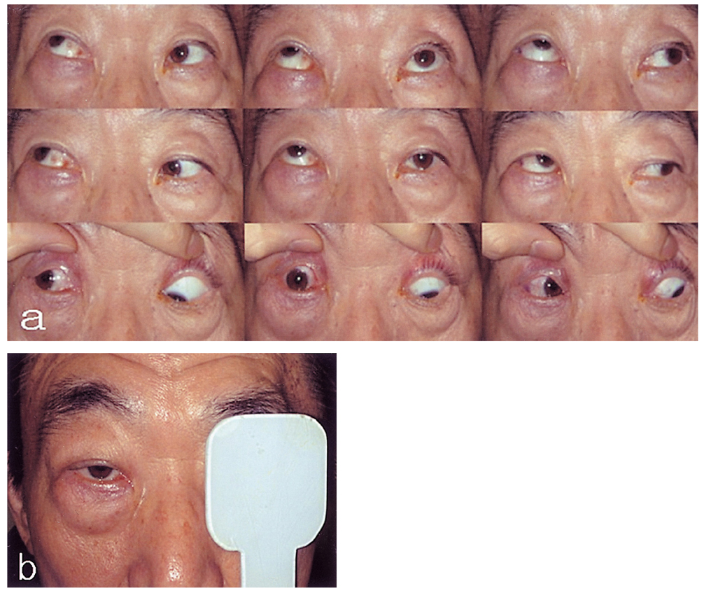
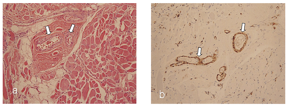
- PURPOSE: To report the case of a patient with large-angle hypertropia of an intramuscular hemangioma of the right superior rectus muscle (SR). METHODS: A 63-year-old man with progressive vertical deviation of the right eye for the past 6 months visited our strabismus department; his condition was not painful. An examination indicated that he had 60PD of right hypertropia at distance and near in primary gaze. Additionally, a significant limitation of his downgaze was noted. The right eye appeared mildly proptotic, and the upper and lower eyelids were slightly edematous. Corrected vision was 20/20 in both eyes. RESULTS: Orbital magnetic resonance imaging (MRI) studies revealed fusiform enlargement of the right superior rectus muscle, with prominent but irregular enhancement following gadolinium administration. Incisional biopsy revealed an intramuscular hemangioma in the superior rectus muscle with cavernous-type vessels. CONCLUSIONS: This case demonstrates that intramuscular hemangioma should be considered in the differential diagnosis of isolated extraocular muscle enlargement and unusual strabismus.
MeSH Terms
Figure
Cited by 1 articles
-
Intramuscular Hemangioma Mimicking Myofascial Pain Syndrome : A Case Report
Dong Hwee Kim, Miriam Hwang, Yoon Kyoo Kang, In Jong Kim, Yoon Kun Park
J Korean Med Sci. 2007;22(3):580-582. doi: 10.3346/jkms.2007.22.3.580.
Reference
-
1. Lee HC, Lee SJ, Kim YD. Intramuscular hemangioma of the upper lid. J Korean Ophthalmol Soc. 2003. 44:2428–2433.2. Wolf GT, Daniel F, Krause CJ, Kaufman RS. Intramuscular hemangioma of the head and neck. Laryngoscope. 1985. 95:21–23.3. Rossiter JL, Hendrix RA, Tom LWC, Potsic WP. Intramuscular hemangioma of the head and neck. Laryngoscope. 1993. 108:18–26.4. Christensen SR, Borgesen SE, Heegaard S, Prause JU. Orbital Intramuscular hemangioma. Acta Ophthalmol Scand. 2002. 80:336–339.5. Kiratli H, Bilgic S, Caglar M, Soylemezoglu F. Intramuscular hemangioma of extraocular muscles. Ophthalmology. 2003. 110:564–568.6. Allen PW, Enzinger FM. Hemangioma of skeletal muscle: An analysis of 89 cases. Cancer. 1972. 29:8–22.7. Trokel SL, Hilal SK. Recognition and differential diagnosis of enlarged extraocular muscles in computed tomography. Am J Ophthamol. 1979. 87:503–512.8. Char DH, Miller T, Kroll S. Orbital metastases: diagnosis and course. Br J Ophthalmol. 1997. 81:386–390.9. Hornblass A, Jacobiec FA, Reifler DM, Mines J. Orbital lymphoid tumors located predominately within extraocular muscles. Ophthalmology. 1987. 94:688–697.10. Buetow PC, Kransdorf MJ, Moser RP, et al. Radiologic appearance of intramuscular hemangioma with emphasis on MR imaging. Am J Radiol. 1990. 154:563–567.11. Kushner BJ. Infantile orbital hemangioma. Int Pediatr. 1990. 5:249–257.
- Full Text Links
- Actions
-
Cited
- CITED
-
- Close
- Share
- Similar articles
-
- Incidentally Found Intramuscular Hemangioma, Mimicking Traumatic Hematoma after Military Training: A Case Report
- Cavernous Hemangioma of the Masseter Muscle
- Intramuscular hemangioma formation in the masseter muscle: a case report
- Intramuscular Hemangioma of the Mentalis Muscle: A Case Report
- A Case of Intramuscular Muller Muscle Hemangioma of Upper Eyelid Mimicking Sarcoidosis


