Korean J Ophthalmol.
2005 Sep;19(3):208-212. 10.3341/kjo.2005.19.3.208.
Investigation of Hemodynamic Changes in the Ophthalmic Artery using Color Doppler Imaging after Strabismus Surgery
- Affiliations
-
- 1Department of Ophthalmology, Chungnam National University College of Medicine, Daejeon, Korea. irismd@cnuh.co.kr
- KMID: 754426
- DOI: http://doi.org/10.3341/kjo.2005.19.3.208
Abstract
- PURPOSE
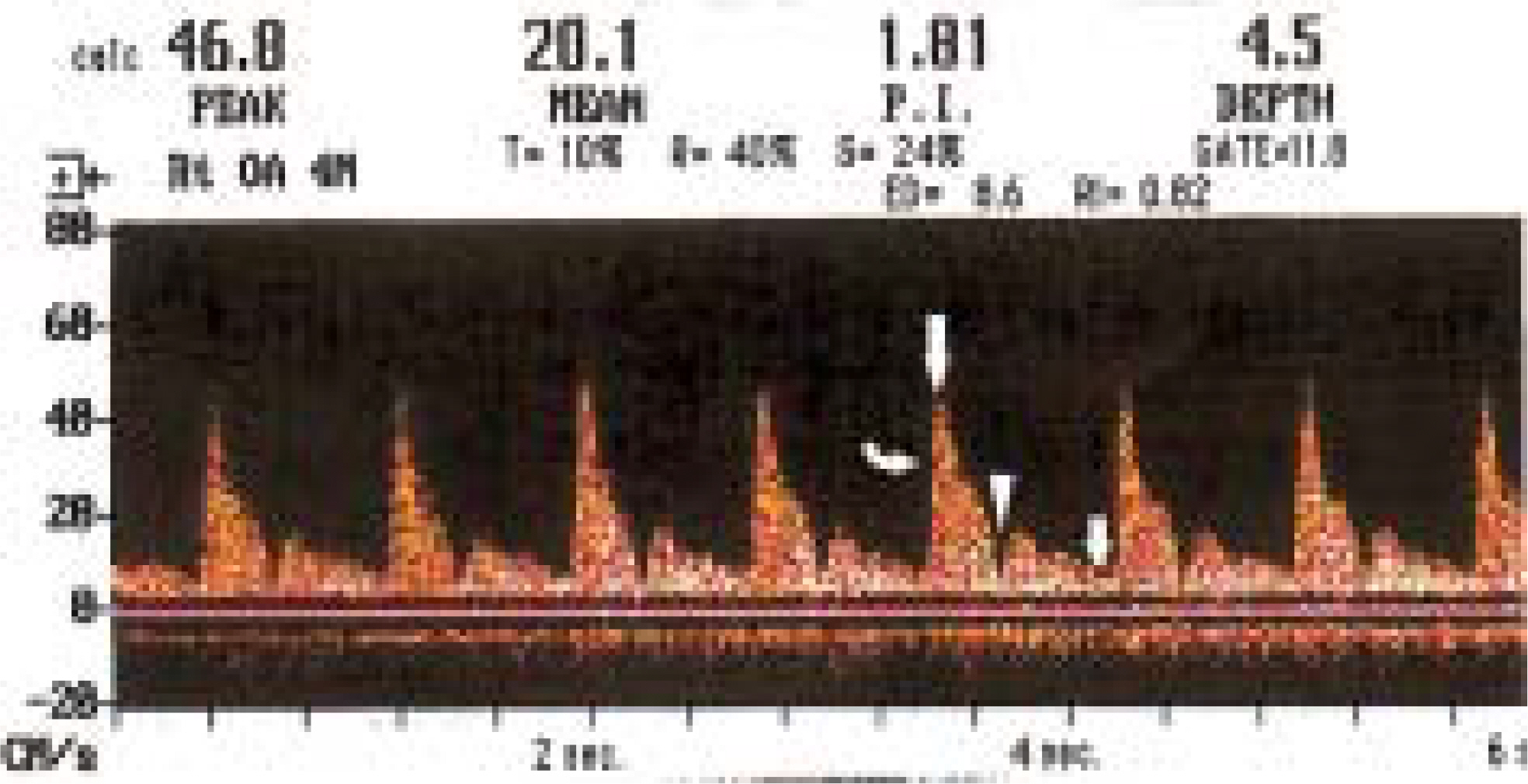
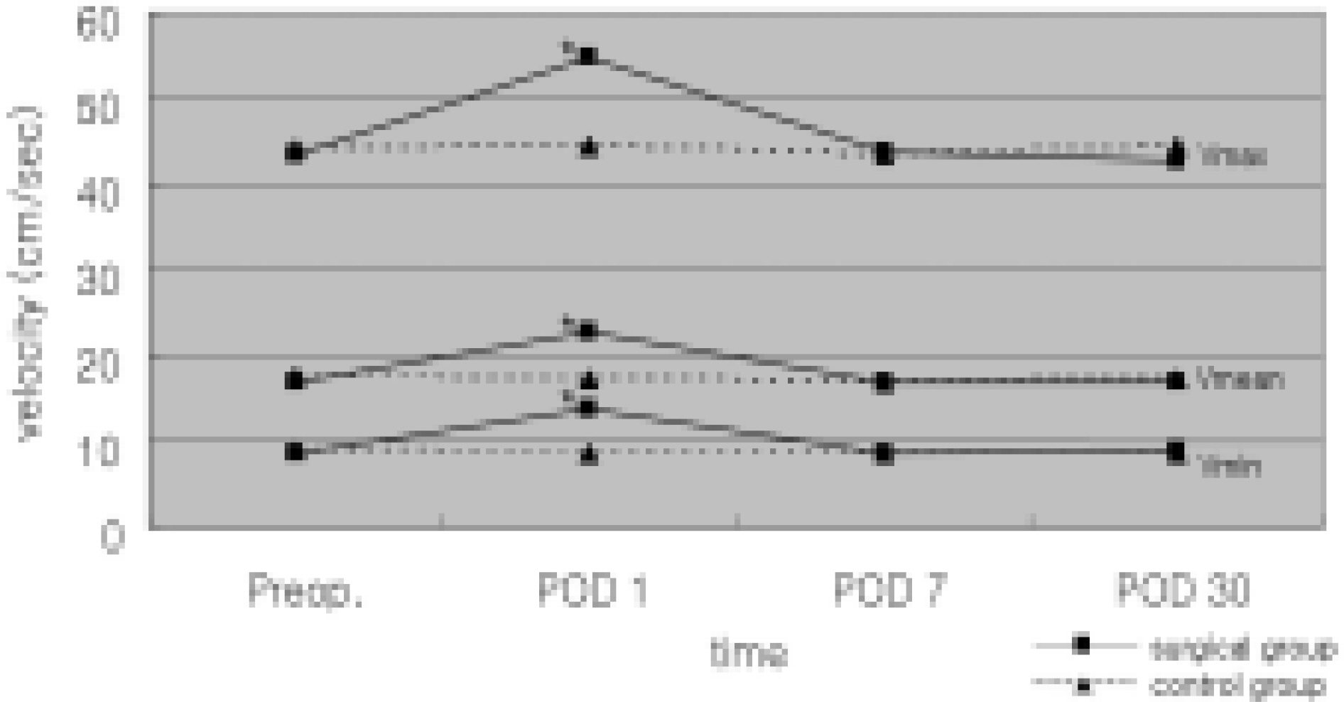
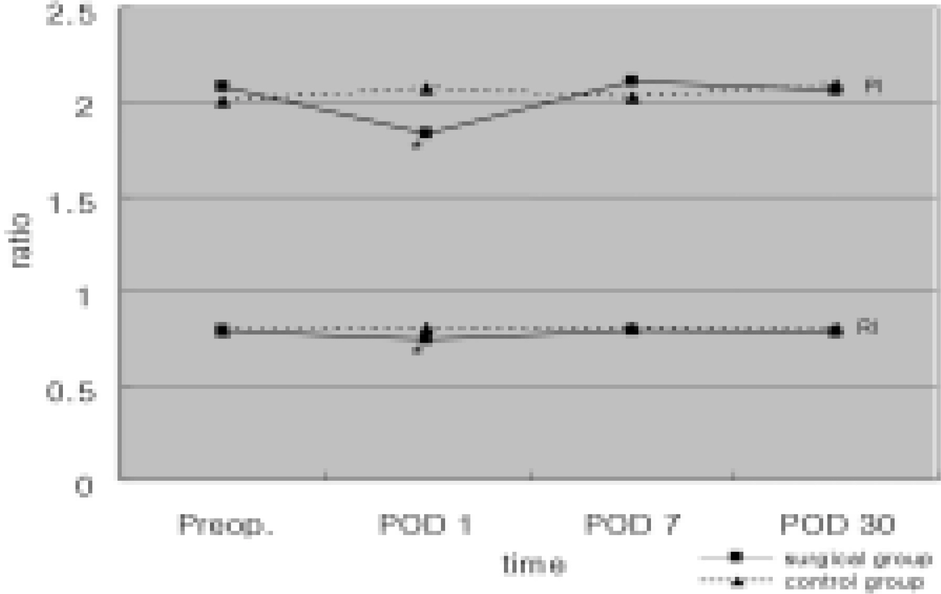
We investigated hemodynamic changes in the ophthalmic artery (OA) using color Doppler imaging (CDI) after two horizontal rectus muscles surgery. METHODS: Eyes of the surgical group (n=18) underwent surgery on two horizontal rectus muscles, and the control group was the contralateral eyes. CDI of the OA was performed before operation and on postoperative days (POD) 1, 7 and 30. Peak systolic (Vmax), end diastolic (Vmin), and mean (Vmean) blood flow velocities were measured, and resistivity index (RI) and pulsatility index (PI) were calculated. RESULTS: Vmax, Vmin and Vmean were significantly higher, and RI and PI were significantly lower in the surgical group than in the control group on POD 1 (p< 0.05). In the surgical group, Vmax, Vmin and Vmean were significantly higher, and RI and PI were significantly lower, on POD 1 than those mesured on other days (p< 0.05). CONCLUSIONS: We showed that surgery on the two horizontal rectus muscles increased OA blood flow during the early postoperative period.
Keyword
MeSH Terms
Figure
Reference
-
1. Erickson SJ, Hendrix LE, Massaro BM, et al. Color Doppler flow imaging of the normal and abnormal orbit. Radiology. 1989; 173:511–6.
Article2. Langham ME, Farrell RA, O'Brien V, et al. Blood flow in the human eye. Acta Ophthalmol. 1989; 191:9–13.
Article3. Guthoff RF, Berger RW, Winkler P, et al. Doppler ultrasonography of the ophthalmic and central retinal vessels. Arch Ophthalmol. 1991; 109:532–6.
Article4. Regillo CD, Sergott RC, Brown GC. Successful scleral buckling procedures decrease central retinal artery blood flow velocity. Ophthalmology. 1993; 100:1044–9.
Article5. Santos L, Carpeans C, Gonzalez F. Ocular blood flow velocity reduction after buckling surgery. Graefes Arch Clin Exp Ophthalmol. 1994; 232:666–9.
Article6. Trible JR, Sergott RC, Spaeth GL, et al. Trabeculectomy is associated with retrobulbar hemodynamic changes. A color Doppler analysis. Ophthalmology. 1994; 101:340–51.7. Mittra RA, Sergott RC, Flaharty PM, et al. Optic nerve decompression improves hemodynamic parameters in papilledema. Ophthalmology. 1993; 100:987–97.
Article8. Lee JP, Olver JM. Anterior segment ischemia. Eye. 1990; 4:1–6.9. Aburn NS, Sergott RC. Orbital colour Doppler imaging. Eye. 1993; 7:639–47.
Article10. Virdi PS, Heyreh SS. Anterior segment ischemia after recession of various recti. An experimental study. Ophthalmology. 1987; 94:1258–71.11. Wilcox LM, Keough EM, Connolly RJ, Hotte CE. The contribution of blood flow by anterior ciliary arteries to the anterior segment in the primate eye. Exp Eye Res. 1980; 30:167–74.12. Mckeown CA, Lambert HM, shore JW. Preservation of anterior ciliary vessels during extraocular muscle surgery. Ophthalmology. 1989; 96:498–506.13. The Korean Strabismus and Pediatric Ophthalmological Society. Gross anatomy of the extraocular muscles. In: Current Concepts in Strabismus. 1st ed.Reston: Naewae Haksool;2004. p. 16–7.14. Hayreh SS. Proceedings: Anatomy and pathophysiology of ocular circulation. Exp Eye Res. 1973; 17:387–8.15. France TD, Simon JW. Anterior segment ischemia syndrome following muscle surgery; the AAPOS experience. J Pediatr Ophthalmol Strabismus. 1986; 23:87–91.
Article16. Elsas FJ, Witherspoon CD. Anterior segment ischemia after strabismus surgery in child. Am J Ophthalmol. 1987; 6:833–4.17. Hiatt RL. Production of anterior segment ischemia. Trans Am Ophthalmol Soc. 1977; 75:87–102.
Article18. Saunders RA, Sandall GS. Anterior segment ischemia syndrome following rectus muscle transposition. Am J Ophthalmol. 1982; 93:34–8.
Article19. Simon JW, Price EC, Krohel GB, et al. Anterior segment ischemia following strabismus surgery. J Pediatr Ophthalmol Strabismus. 1984; 21:179–84.
Article20. Wagner RS, Nelson LB. Complications following strabismus surgery. Int Ophthalmol Clin. 1985; 25:171–8.
Article21. Hayreh SS. Scott WE. Fluorescein iris angiography. II. Disturbances of iris circulation following strabismus operation on the various recti. Arch Ophthalmol. 1978; 96:1390–400.22. Hayreh SS. Scott WE. Fluorescein iris angiography. I. Normal pattern. Arch Ophthalmol. 1978; 96:1383–9.23. Meyer PA, Watson PG. Low dose fluorescein angiography of the conjunctiva and episclera. Br J Ophthalmol. 1987; 71:2–10.
Article24. Ormerod LD, Fariza E, Hughes GW, et al. Anterior segment fluorescein videoangiography with a scanning angiographic microscope. Ophthalmology. 1990; 97:745–51.
Article25. Lieb WE, Cohen SM, Merton DA, et al. Color Doppler imaging of the eye and orbit: technique and normal vascular anatomy. Arch Ophthalmol. 1991; 109:527–31.26. Saliba E, Laugier J. Circulation du nouveau-ne' et de l'enfant. Pourcelot L, editor. Dynamique Cardio-Vasculaire Foetale et Neonatale Echocardiographie-Doppler. 1st ed.Paris: Masson;2006. p. 139–61.27. Freed KS, Brown CK, Carrol BA. The extracranial cerebral vessels. Rumack CM, Wilson SR, Charboneau JW, editors. Diagnostic Ultrasound. 1st ed.St Louis: Mosby;2006. p. 26–30.28. Peter AN, Scott WS, Alon H. Color Doppler ultrasound measurements after topical and retrobulbar epinephrine in primate eyes. Invest Ophthalmol Vis Sci. 1997; 38:2655–61.29. Pelit A, Barutcu O, Oto S, Aydin P. Investigation of hemodynamic changes after strabismus surgery using color Doppler imaging. J AAPOS. 2002; 6:224–7.
Article30. Bayramlar H, Totan Y, Cekic O, et al. Evaluation of hemodynamic changes in the ophthalmic artery with color Doppler ultrasonography after strabismus surgery. J Pediatr Ophthalmol Strabismus. 2000; 37:94–100.
Article31. Tane S, Hashimoto T. Estimation of blood flow in the carotid artery and intraorbital ophthalmic artery by color pulse Doppler ultra sonography. Acta Ophthalmol. 1992; 204:S62–5.32. Han KH, Kim BJ, Youn JW, Lee HB. The measurement of ocular blood flow velocity using Doppler ultrasound in normal eyes. J Korean Ophthalmol Soc. 1994; 35:1685–90.33. Hong JW, Kim YY, Lee TS. Measurement of blood flow velocity of ophthalmic and central retinal artery using color Doppler imaging. J Korean Ophthalmol Soc. 1996; 37:985–92.34. Goebel W, Lieb W, Ho A, et al. Color Doppler Imaging: A new technique to assess orbital blood flow in patients with diabetic retinopathy. Invest Ophthalmol Vis Sci. 1995; 36:864–70.35. Lizzi FL, Mortimer AJ. Bioeffects considerations for the safety of diagnostic ultrasound. J Ultrasound Med. 1988; 7:S1–38.36. Lizzi FL, Packer AJ, Coleman DJ. Experimental cataract production by high frequency ultrasound. Ann Ophthalmol. 1978; 10:934–42.37. Lizzi FL, Coleman DJ, Driller J, et al. Effects of pulsed ultrasound on ocular tissue. Ultrasound Med Biol. 1981; 7:245–52.
Article
- Full Text Links
- Actions
-
Cited
- CITED
-
- Close
- Share
- Similar articles
-
- Measurement of Blood Flow Velocity of Ophthalmic and Central Retinal Artery using Color Doppler Imaging
- Color Doppler Imaging to Diagnose Ocular Disorders
- Color Doppler Imaging of Subclavian Steal Phenomenon
- Color Doppler Ultrasonography in the Evaluation of the Acute Scrotum
- Color Doppler Velocimetry in Primary Open Angle Glaucoma


