Korean J Ophthalmol.
2005 Jun;19(2):149-152. 10.3341/kjo.2005.19.2.149.
Development of Pupillography Using Image Processing
- Affiliations
-
- 1Department of Biomedical Engineering, Seoul National University College of Medicine, Seoul, Korea.
- 2SLMED, Seoul, Korea.
- 3Seoul National University College of Medicine, Seoul National University Bundang Hospital, Seongnam, Korea. hjm@snu.ac.kr
- KMID: 754415
- DOI: http://doi.org/10.3341/kjo.2005.19.2.149
Abstract
- PURPOSE
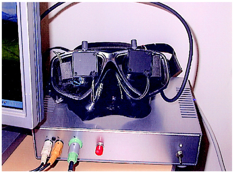
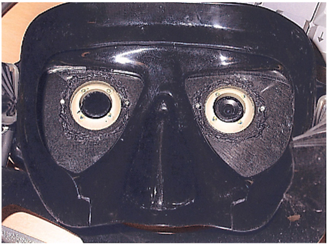
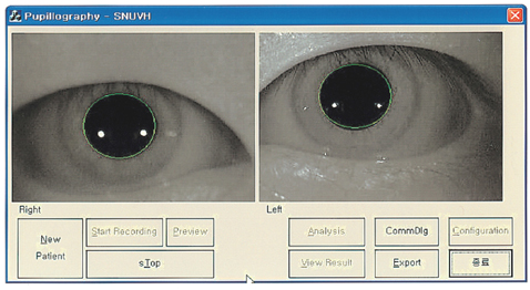
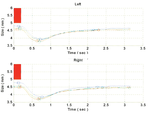
Pupillary examination is an important objective method to diagnose lesions of the anterior visual pathways. However, errors and faults may easily alter the interpretation and value of the test as it is highly dependent on the examiner's skills. Therefore, we tried to develop a pupillography which is independent of the examiner. METHODS: Hardware composed of a binocularly measuring instrument adapted for an infrared charge coupled device (CCD) was developed. Two arrays of infrared light emitting diodes (LED) were supplied in front of each of the subject's eyes. A microcontroller to modulate these LED was developed, as was software to save and analyze the pupil images. The hardware was able to deliver a light to either eye or to both eyes, and to change the time, frequency, and intensity of the stimulus. The software automatically analyzed the pupil size and location by image processing. Pupil size was calculated continuously. After artifact elimination, the response amplitudes of the pupils were determined for the right and left pupils. RESULTS: Pupillary images of size 320 x 240, at 30 frames/second, were saved and processed to evaluate the change of the actual pupil size and the velocity of pupillary response. CONCLUSIONS: A pupillography to measure, save and analyze the pupillary response using image processing was developed. Further detailed clinical studies with a large number of patients will be required to validate this new method.
Keyword
MeSH Terms
Figure
Reference
-
1. Levatin P. Pupillary escape in disease of the retinal or optic nerve. Arch Ophthalmol. 1959. 62:768–779.2. Wilhelm H. Pupil examination and evaluation of pupillary disorders. Neuroophthalmology. 1994. 14:283–295.3. Wilhelm H, Wilhelm B. Clinical applications of pupillography. J Neuroophthalmol. 2003. 23:42–49.4. Wilhelm H. Neuro-ophthalmology of pupillary function-practical guidelines. J Neurol. 1998. 245:573–583.5. Uren SM, Stewart P, Crosby PA. Subject cooperation and the visual evoked response. Invest Ophthalmol Vis Sci. 1979. 18:648–652.6. Thompson HS, Montague P, Cox TA, Corbett JJ. The relationship between visual acuity, pupillary defect, and visual field loss. Am J Ophthalmol. 1982. 93:681–688.7. Kaback MB, Burde RM, Becker B. Relative afferent pupillary defect in glaucoma. Am J Ophthalmol. 1976. 81:462–468.8. Firth AY. Pupillary responses in amblyopia. Br J Ophthalmol. 1990. 74:676–680.9. Lowenstein O. Clinical pupillary symptoms in lesions of the optic nerve, optic chiasm and optic tract. Arch Ophthalmol. 1954. 52:385–403.10. Lowenstein O, Loewenfeld IE. Electronic pupillography: a new instrument and some clinical applications. Arch Ophthalmol. 1958. 59:352–363.11. Thompson HS. Afferent pupil defects. Pupillary findings associated with defects of the afferent arm of the pupillary light reflex arc. Am J Ophthalmol. 1966. 62:860–873.12. Fison PN, Garlick DJ, Smith SE. Assessment of unilateral afferent pupillary defects by pupillography. Br J Ophthalmol. 1979. 63:195–199.13. Cox TA. Pupillography of a relative afferent pupillary defect. Am J Ophthalmol. 1986. 101:320–324.


