Granulosa Cell Tumor of Scrotal Tunics: A Case Report
- KMID: 754115
- DOI: http://doi.org/10.3348/kjr.2001.2.2.117
Abstract
- We report a case of adult granulosa cell tumor arising in the scrotal tunics. The patient was a 34-year-old man who presented with right scrotal swelling, first noticed four months previously. Under the initial clinical impression of epididymo-orchitis, antibiotic treatment was instituted but there was no response. The paratesticular nodules revealed by ultrasound and magnetic resonance imaging mimicked intratesticular lesion, and radical orchiectomy was performed. Although several cases of adult testicular granulosa cell tumor, have been reported, the occurrence of this entity in the paratesticular area has not, as far as we are aware, been previously described.
Keyword
MeSH Terms
Figure
-
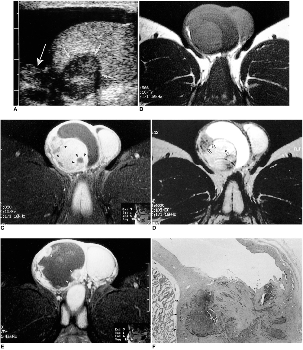
Fig. 1 Axial MR images of the scrotum. A. Sagittal sonogram of the right side of the scrotum. A well-defined hypoechoic mass (short arrows) in the tesitis and a small hypoechoic nodule adjacent to the upper pole of the testis (long arrow) are seen. Extensive hydrocele is present. B, C. T1-weighted (A), Gd-enhanced T1-weighted (B), and T2-weighted (C) MR images reveal well-defined nodules in the testis and adjacent to it. In image B, strong peripheral enhancement is observed (arrowheads). Note that on the T2-weighted image there is a dark rim that separates normal testis from the nodule (arrows). E. In the lower portion of the scrotum there is extensive hydrocele, and small enhancing nodules (arrowheads) are seen along the inner margin of the scrotal wall. F. Pathologic findings of the tumor. Histopathologic specimen shows a nodule between the tunica vaginalis and the testis. Note the preservation of the inner margin of the tunica albuginea (arrows) (H and E, original magnification ×40).
Reference
-
1. Hricak H, Hamm B, Kim B. The epididymis, spermatic cord, and paratesticular tissue: congenital anomalies and tumors. Imaging of the scrotum. 1995. New York: Raven Press;129.2. Makarainen H, Tammela T, Karttunen T, et al. Intrascrotal adenomatoid tumors and their ultrasound findings. J Clin Ultrasound. 1993. 21:33–37.3. Holden A, List A. Extratesticular lesions: a radiological and pathological correlation. Australas Radiol. 1994. 38:99–105.4. Yeager B, Arger P, Mintz M, et al. The impact of sonography on the management of extratesticular abnormalities of the scrotum. J Clin Ultrasound. 1989. 17:573–577.5. Vick C, Bird K, Rosenfield A, Viscomi G, Taylor K. Scrotal masses with uniformly hyperechoic pattern. Radiology. 1983. 148:209–211.6. Haller J, Tscholakoff D, Gundry C, et al. Sonography of unusual extratesticular lesions. Urol Radiol. 1989. 11:190–193.7. Frates MC, Benson CB, DiSalvo DN, et al. Solid extratesticular masses evaluated with sonography: pathologic correlation. Radiology. 1997. 204:33–37.8. Beccia D, Krane R, Olsson C. Clinical management of non-testicular intrascrotal tumors. J Urol. 1976. 116:476–479.9. Maurer R, Taylor CR, Schmucki O, Hedinger CE. Extratesticular gonadal stromal tumor in the pelvis: A case report with immunoperoxidase findings. Cancer. 1980. 45:985–990.10. Jimenez-Quintero LP, Ro JY, Zavala-Pompa A, et al. Granulosa cell tumor of the adult testis: a clinicopathologic study of seven cases and a review of the literature. Hum Pathol. 1993. 24:1120–1125.11. Nistal M, Lazaro R, Garcia J, Paniagua R. Testicular granulosa cell tumor of the adult type. Arch Pathol Lab Med. 1992. 116:284–287.12. Gaylis FD, August C, Yeldandi A, Nemcek A, Garnett J. Granulosa cell tumor of the testis: Ultrastructural and ultrasonographic characteristics. J Urol. 1989. 141:126–127.13. McCluggage WG, Shanks JH, Whiteside C, et al. Immunohistochemical study of testicular sex cord-stromal tumors, including staining with anti-inhibin antibody. Am J Surg Pathol. 1998. 22(5):615–619.14. Feimanis MG, Bohm-Velez M, Mendelson EB, Erickson ER, Campanella SD. Ultrasound appearanace of extratesticular myosarcoma. J Clin Ultrasound. 1991. 19:101–104.
