Foreign Body Granulomas of the Breast Presenting as Bilateral Spiculated Masses
- KMID: 754114
- DOI: http://doi.org/10.3348/kjr.2001.2.2.113
Abstract
- In Asia, mammography following the injection of foreign materials into the breasts for cosmetic augmentation is frequently seen and diagnosis based on the typical radiologic findings is straightforward. We report the unusual radiologic findings in two patients with foreign body granulomas caused by injected foreign materials and discovered incidentally during screening work up. The mammographic findings were bilateral, hyperdense, spiculated masses, with occasional microcalcification, and at sonography, markedly hypoechoic, spiculated solid masses, located near the pectoralis muscle and partly extending into it, were observed. These radiologic findings mimicked malignancy.
Keyword
MeSH Terms
Figure
-
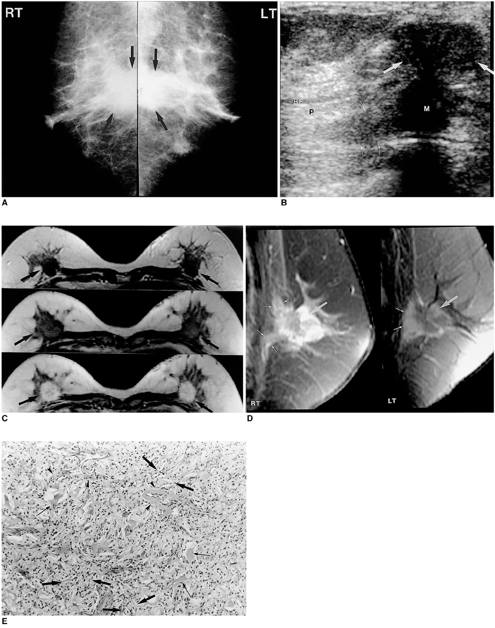
Fig. 1 61-year-old woman with foreign body granuloma of the breast. A. Bilateral mediolateral oblique mammograms show ill-defined masses with some spiculation in the central posterior portion of both breasts (arrows). B. Sonogram of the right breast shows a markedly lobulated, heterogeneously hypoechoic mass in the subareolar portion (large arrows) and extending into the pectoralis muscle (small arrows). A sonogram of the left breast revealed similar features (not shown) (M: retroglandular portion of the mass; P: pectoralis muscle; RF: retromammary fat). C. Axial, fat-saturated, T2-weighted fast spin-echo images (TR/TE, 4383/102, upper), two-dimensional spoiled gradient-echo images before (76/3.3, middle) and 1 min aftar gadolinium enhancement (lower) show bilateral, spiculated, low-signal intensity masses with early enhancement (arrows) in the posterior central portion of the breasts (arrows). D. Sagittal T1-weighted images with fat saturation 8 mins after gadolinium enhancement (550/10) show spiculated masses in both breasts (large arrows) extending into the pectoralis muscle (small arrows). Note the delayed peripheral enhancement of the masses. E. Photomicrograph of histologic specimen shows cholesterol clefts, seen as needle-like empty spaces (large arrows), secretory materials in luminal structures (small arrows) and perivascular fibrosis (arrowheads) (H and E, ×100).
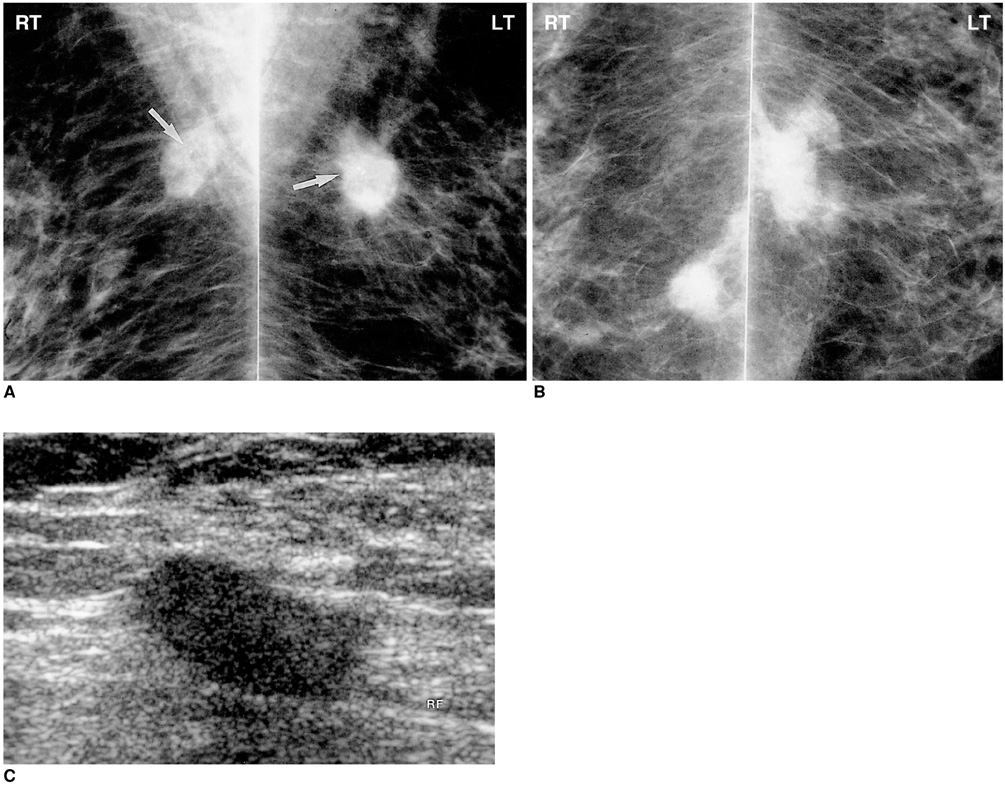
Fig. 2 64-year-old woman with foreign body granuloma of the breast. A, B. Bilateral mediolateral oblique (A) and craniocaudal (B) mammograms show high-density spiculated masses in the posterior portion of each breast. Microcalcifications are seen within the masses (arrows). C. Sonogram of the left breast reveals an ovoid hypoechoic mass with suspicious fine spiculation at its posterior portion (RF: retromammary fat).
Reference
-
1. Kopans DB. Breast Imaging. 1997. 2nd ed. Philadelphia: Lippincott-Raven;302–305.2. Rosenberg RF, Siegelman SS. Foreign-body granuloma simulating carcinoma on mammography. N Y State J Med. 1976. 76:445–446.3. Wakabayashi M, Reid JD, Bhattacharjee M. Foreign body granuloma caused by prior gunshot wound mimicking malignant breast mass. AJR. 1999. 173:321–322.4. Patrikeos A, Wylie EJ, Bourke A, Frost F. Imaging of carbon granulomas of the breast following carbon track localization. Clin Radiol. 1998. 53:845–848.5. Helbkch TH, Wunderbaldinger P, Plenk H, et al. The value of MRI in silicone granuloma of the breast. European J Radiol. 1997. 24:155–158.6. Khong PL, Ho LWC, Chan JHM, Leong LLY. MR imaging of breast paraffinomas. AJR. 1999. 173:929–932.7. Som PM, Curtin HD, editors. Head and Neck Imaging. 1996. 3rd ed. St. Louis: Mosby-Year Book;1396–1398.8. Wilhelmus JL, Schrodt GR, Mahaffey LM. Cholesterol granulomas of the breast : a lesion which clinically mimics carcinoma. Am J Clin Pathol. 1982. 77:592–597.9. Reynolds HE, Crame HM. Cholesterol granuloma of the breast: a mimic of carcinoma. Radiology. 1994. 191:249–250.10. Newstead GM, Weinreb JC. Critical pathways for the future: MR imaging and digital mammography. RadioGraphics. 1995. 15(4):951–962.11. Dao TH, Rahmouni A, Campana F, et al. Tumor recurrence versus fibrosis in the irradiated breast: differentiation with dynamic gadolinium-enhanced MR imaging. Radiology. 1993. 187(3):751–755.
- Full Text Links
-
- Actions
-
Cited
- CITED
-
- Close
- Share
-
- Similar articles
-
- Immediate Breast Reconstruction Using Bilateral Free TRAM Flap after Removal of Paraffinoma
- Foreign Body Granulomas after the Use of Dermal Fillers: Pathophysiology, Clinical Appearance, Histologic Features, and Treatment
- Localized Foreign Body Granulomas of the Breast: Clinical and Mammographic Findings
- Clinical Observation on Sperm Granulomas after Vasectomy
- Facial Foreign Body Granulomas Caused by Filler Injection and Barbed Thread-lifting
