Giant Serpentine Intracranial Aneurysm: A Case Report
- Affiliations
-
- 1Department of Radiology, Wonju College of Medicine, Yonsei University Wonju Christian Hospital, Kangwon-do, Korea. cursor2@wonju.yonsei.ac.kr
- KMID: 754105
- DOI: http://doi.org/10.3348/kjr.2001.2.3.179
Abstract
- The authors present a case of giant serpentine aneurysm (a partially thrombosed aneurysm containing tortuous vascular channels with a separate entrance and outflow pathway). Giant serpentine aneurysms form a subgroup of giant intracranial aneurysms, distinct from saccular and fusiform varieties, and in this case, too, the clinical presentation and radiographic features of CT, MR imaging and angiography were distinct.
MeSH Terms
Figure
-
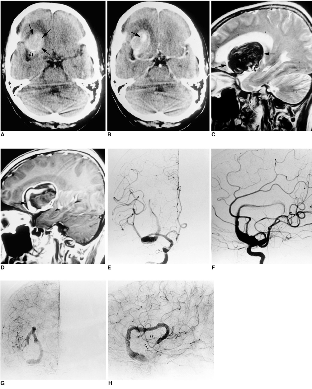
Fig. 1 A 50-year-old woman with giant intracranial aneurysm. A. Precontrast CT scan shows a large oval-shaped mass with increased attenuation representing thrombus (arrows) and slight peripheral calcification (arrowheads) in the right frontal region. Peripheral edema may also be observed. B. Postcontrast CT scan demonstrates peripheral and central enhancenment. The enhanced central tubular structure represents the serpentine vascular channel (arrow). C. T2-weighted sagittal image shows a large thrombosed aneurysm (arrows) with peripheral edema (arrowheads) in the right frontal area. D. Postcontrast T1-weighted sagittal image shows an enhancing tubular structure (arrow) indicating the serpentine vascular channel. E, F. Angiograms of the right internal carotid artery. Anteroposterior (E) and lateral (F) arterial-phase angiograms show the ectatic vascular channel involving the upper trunk of the right middle cerebral artery and corresponding to the vascular channel identified on MR images. G, H. Anteroposterior (G) and lateral (H) capillary phase angiograms more clearly demonstrate the tortuous vascular channel of the serpentine aneurysm and show delayed filling of normal cortical branches of the superior division (arrowheads).
Cited by 1 articles
-
Endovascular Treatment of Giant Serpentine Aneurysm of the Middle Cerebral Artery
Young Ha Jeong, Jong Yeon Kim, Youn Moo Koo, Jong Wook Choi, Kum Whang, Chul Hu, Sung Min Cho
J Cerebrovasc Endovasc Neurosurg. 2016;18(3):264-270. doi: 10.7461/jcen.2016.18.3.264.
Reference
-
1. Segal HD, McLaurin RL. Giant serpentine aneurysm: Report of two cases. J Neurosurg. 1977. 46:115–120.2. Aletich VA, Debrun GM, Monsein LH, Nanta HJW, Spetzler RF. Giant serpentine aneurysms: a review and presentation of five cases. Am J Neuroradiol. 1995. 16:1061–1072.3. Tomasello F, Albanese V, Cioffi FA. Giant serpentine aneurysms: a separate entity. Surg Neurol. 1979. 12:429–432.4. Fodstad H, Liliequist B, Wirell S, Nilsson PE, Boquist L, Abdul-Rahman A. Giant serpentine intracranial aneurysm after carotid ligation: case report. J Neurosurg. 1978. 49:903–909.5. Haddad GF, Haddad FS. Cerebral giant serpentine aneurysm: case report and review of the literature. Neurosurgery. 1988. 23:92–97.6. Belec L, Cesaro P, Brugieres P, Gray F. Tumor-simulating giant serpentine aneurysm of the posterior cerebral artery. Surg Neurol. 1988. 29:210–215.7. Vishteh AG, Spetzler RF. Evolution of a dolichoectatic aneurysm into a giant serpentine aneurysm during long-term follow up: case illustration. J Neurosurg. 1999. 91:346.
- Full Text Links
-
- Actions
-
Cited
- CITED
-
- Close
- Share
-
- Similar articles
-
- Giant Serpentine Intracranial Aneurysm Treated with Wrapping under the Extracorporeal Circulation and Hypothermia
- Giant Serpentine Aneurysm of the Middle Cerebral Artery
- Giant Serpentine Aneurysm of the Anterior Communicating Artery: Case Report
- Giant Serpentine Aneurysm of the Posterior Cerebral Artery: Case Report
- Endovascular Treatment of Giant Serpentine Aneurysm of the Middle Cerebral Artery
