Korean J Radiol.
2001 Sep;2(3):132-137. 10.3348/kjr.2001.2.3.132.
Esophageal Leiomyoma: Radiologic Findings in 12 Patients
- Affiliations
-
- 1Department of Radiology Samsung Medical Center, Sungkyunkwan University School of Medicine, Seoul, Korea. kslee@smc.samsung.co.kr
- KMID: 754097
- DOI: http://doi.org/10.3348/kjr.2001.2.3.132
Abstract
OBJECTIVE
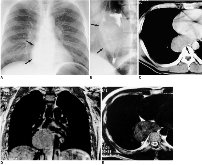
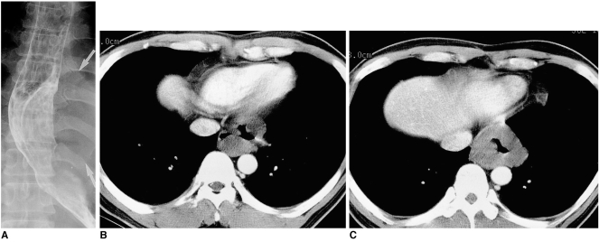
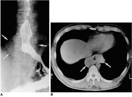
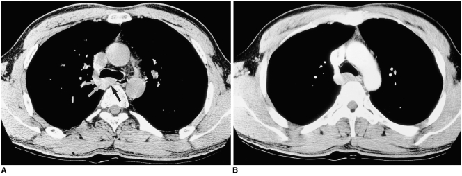
The aim of our study was to describe and compare the radiologic findings of esophageal leiomyomas. MATERIALS AND METHODS: The chest radiographic (n = 12), esophagographic (n = 12), CT (n = 12), and MR (n = 1) findings of surgically proven esophageal leiomyomas in 12 consecutive patients [ten men and two women aged 34 - 47 (mean, 39) years] were retrospectively reviewed. RESULTS: The tumors, surgical specimens of which ranged from 9 to 90 mm in diameter, were located in the upper (n = 1), middle (n = 5), or lower esophagus (n = 6). In ten of the 12 patients, chest radiography revealed the tumors as mediastinal masses. Esophagography showed them as eccentric, smoothly elevated filling defects in 11 patients and a multilobulated encircling filling defect in one. In 11 of the 12 patients, enhanced CT scans revealed a smooth (n = 9) or lobulated (n = 2) tumor margin, and attenuation was homogeneously low (n = 7) or iso (n = 4). In one patient, the tumor signal seen on T2-weighted MR images was slightly high. CONCLUSION: Esophageal leiomyomas, located mainly in the middle or distal esophagus, are consistently shown by esophagography to be mainly eccentrically elevated filling defects and at CT, lesions showing homogeneous low or isoattenuation are demonstrated.
MeSH Terms
Figure
Reference
-
1. Seremetis MG, Lyons WS, Deguzman VC, Peabody JW. Leiomyomata of the esophagus. Cancer. 1976; 38:2166–2175. PMID: 991129.2. Moersch HJ, Harrington SW. Benign tumor of the esophagus. Ann Otol. 1944; 53:800–817.3. Barreiro F, Seco JL, Molina J, et al. Giant esophageal leiomyoma with secondary megaesophagus. Surgery. 1976; 79:436–439. PMID: 1257906.4. Gallinger S, Steinhardt MI, Goldger M. Giant leiomyoma of the esophagus. Am J Gastroenterol. 1983; 78:708–711. PMID: 6637958.5. Montesi A, Pesaresi A, Graziani L, Salmistraro D, Dini L, Bearzi I. Small benign tumors of the esophagus: radiological diagnosis with double-contrast examination. Gastrointest Radiol. 1983; 8:207–212. PMID: 6618085.
Article6. Megibow AJ, Balthazar EJ, Hulnick DH, Naidich DP, Bosniak MA. CT evaluation of gastrointestinal leiomyomas and leiomyosarcomas. AJR. 1985; 144:727–731. PMID: 3872029.
Article7. Massari M, Lattuada E, Zappa MA, et al. Evaluation of leiomyoma of the esophagus with endoscopic ultrasonography. Hepatogastroenterology. 1997; 44:727–731. PMID: 9222681.8. Pelot D. Haurich WS, Schaffner F, Berk JE, editors. Anatomy, anomalies, and physiology of the esophagus. Bockus Gastroenterology. 1995. 5th ed. Philadelphia: Saunders;p. 397–411.9. Evans HL. Smooth muscle tumors of the gastrointestinal tract. A study of 56 cases followed for a minimum 10 years. Cancer. 1985; 56:2242–2250. PMID: 4052969.10. Lewis B, Maxfield RG. Leiomyoma of the esophagus: Case report and review of the literature. Int Abstr Surg. 1954; 99:105–128. PMID: 13187207.11. Godard JE, McCranie D. Multiple leiomyomas of the esophagus. AJR. 1973; 117:259–262.
Article12. Levine MS, Buck JL, Pantongrag-Brown L, Buetow PC, Lowry MA, Sobin LH. Esophageal leiomyomatosis. Radiology. 1996; 199:533–536. PMID: 8668807.
Article13. Rabushka LS, Fishman EK, Kulman JE, Hurban RH. Diffuse esophageal leiomyomatosis in a patient with Alport syndrome: CT demonstration. Radiology. 1991; 179:176–178. PMID: 2006273.
Article14. Casillas J, Joseph RC, Guerra JJ. CT appearance of uterine leiomyomas. RadioGraphics. 1990; 10:999–1007. PMID: 2259770.
Article15. Schnall MD. Gore RM, Levine MS, editors. Magnetic resonance imaging of the hollow viscera. Textbook of gastrointestinal radiology. 2000. 2nd ed. Philadelphia: Saunders;p. 86–97.16. Totten RS, Stout AP, Humphreys GH, Moore RL. Benign tumors and cysts of the esophagus. J Thorac Surg. 1953; 25:606–622. PMID: 13062346.
Article17. Storey CF, Adams WC. Leiomyoma of the esophagus: report of four cases and a review of the surgical literature. Am J Surg. 1956; 91:3–23. PMID: 13268774.18. Seremetis MG, Lyons WS, de Guzman VC, et al. Leiomyomata of the esophagus: an analysis of 838 cases. Cancer. 1976; 38:2166–2177. PMID: 991129.19. Seremetis MG, de Guzman VC, Lyons WS, et al. Leiomyoma of the esophagus: a report of 19 surgical cases. Ann Thorac Surg. 1973; 16:308–316. PMID: 4728612.
- Full Text Links
- Actions
-
Cited
- CITED
-
- Close
- Share
- Similar articles
-
- An extremely rare coexistence of achalasia cardia with esophageal leiomyoma: An endoscopic treatment approach
- A Case of Esophageal Leiomyoma Showing High FDG Uptake on F-18 FDG PET
- Subphrenic Esophageal Diverticulum Associated with Esophageal Dysmotility and Leiomyoma
- A Case of Subphrenic Esophageal Diverticulum Associated with Esophageal Leiomyoma
- Esophageal leiomyoma combined with achalasia: report of 1 case


