Radiologic Findings of Multiple Myeloma with Gastric Involvement: A Case Report
- Affiliations
-
- 1Department of Diagnostic Radiology, Chonbuk National University Hospital, Korea. jmsh@chonbuk.ac.kr
- KMID: 754073
- DOI: http://doi.org/10.3348/kjr.2002.3.2.133
Abstract
- We report a case of multiple myeloma with gastric involvement occurring in a patient who underwent an upper gastrointestinal series (UGIS), CT and MRI. UGIS depicted a luminal protruding mass, while contrast-enhanced CT demonstrated marked thickening of the gastric wall, with subtle contrast enhancement. At T1- and T2-weighted MR imaging, the mass showed iso- and intermediate signal intensity, respectively. After the administration of contrast material, subtle homogeneous enhancement was apparent.
MeSH Terms
Figure
-
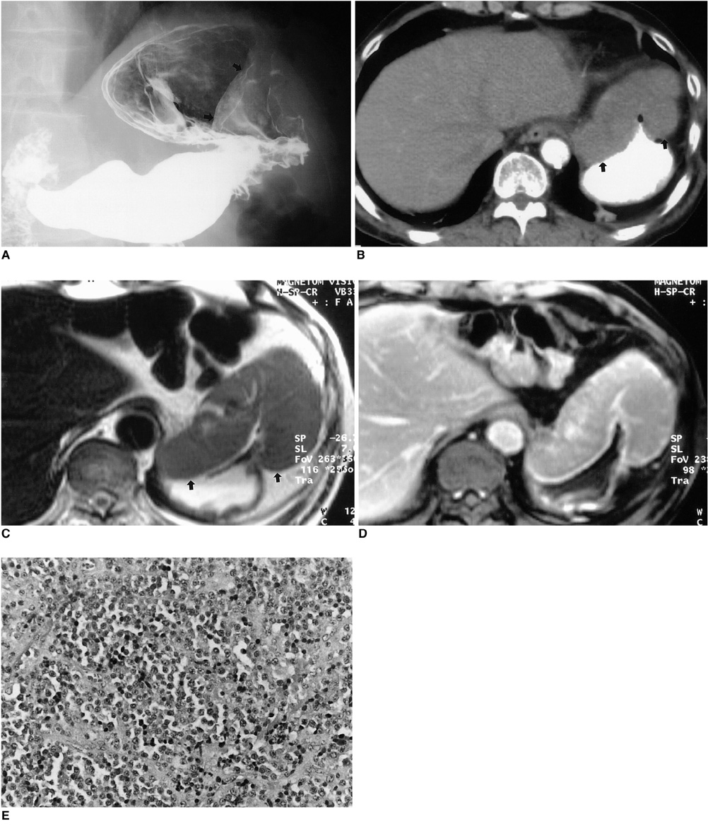
Fig. 1 Multiple myeloma with gastric involvement in a 61-year-old man. A. Upper gastrointestinal series shows a large, protruding, luminal mass (arrows) with a well-defined margin in the anterior wall of the gastric cardia. B. Contrast-enhanced CT scan demonstrates marked gastric wall thickening with subtle contrast enhancement (arrows). C. Axial T2-weighted image shows homogeneous signal intensity slightly higher than that of the liver (arrows). D. Axial gadolinium-enhanced MR image obtained 3 minutes after the administration of contrast material depicts homogeneous enhancement, similar to that of hepatic parenchyma. E. Photograph of endoscopic biopsy specimen shows dense and monotonous infiltration by plasma cells (original magnification, ×200; hematoxylin-eosin staining). Note the presence of a monomorphic population of plasma cells with variable atypia.
Reference
-
1. Kinoshita Y, Watanabe M, Takahashi H, et al. A case of gastric plasmacytoma: genetic analysis and immunofixation electrophoresis. Am J Gastroenterol. 1991. 86:349–353.2. Spagnoli I, Gattoni F, Mazzoni R, Uslenghi C. Primary gastrointestinal plasmacytoma: report of three cases. Diagn Imaging. 1983. 52:23–27.3. Yoon SE, Ha HK, Lee YS, et al. Upper gastrointestinal series and CT findings of primary gastric plasmacytoma: report of two cases. AJR. 1999. 173:1266–1268.4. Pimental RR, Van Stolk R. Gastric plasmacytoma: a rare cause of massive gastrointestinal bleeding. Am J Gastroenterol. 1993. 88:1963–1964.5. Remingio PA, Klaum A. Extramedullary plasmacytoma of the stomach. Cancer. 1971. 27:562–568.6. Marcos HB, Semelka RC. Stomach diseases: MR evaluation using combined T2-weighted single-shot echo train spin-echo and gadolinium-enhanced spoiled gradient-echo sequences. J Magn Reson Imaging. 1999. 10:950–960.
- Full Text Links
-
- Actions
-
Cited
- CITED
-
- Close
- Share
-
- Similar articles
-
- Liver Involvement of Multiple Myeloma Mimicking Intrahepatic Cholangiocarcinoma: A Case Report
- Meningeal and Cerebral Involvement of a Plasmacytoma in an IgG Multiple Myeloma Patient: Case Report
- A Rare Case of Diffuse Pachymeningeal Involvement of Multiple Myeloma
- Multiple Myeloma of the Male Breast: A Case Report
- A Case Report of Extramedullary Myeloma Mimicking Lymphoma with Extensive Abdominal Involvement
