CT and Pathologic Findings of A Case of Subdural Osteoma
- Affiliations
-
- 1Department of Radiology, Seoul Municipal Boramae Hospital, Seoul, Korea.
- 2Department of Pathology, Seoul Municipal Boramae Hospital, Seoul, Korea.
- 3Department of Neurosurgery, Seoul Municipal Boramae Hospital, Seoul, Korea.
- KMID: 754064
- DOI: http://doi.org/10.3348/kjr.2002.3.3.211
Abstract
- A 43-year-old female presented with persistent headache and dizziness which had first occurred two years earlier. The physical and neurological findings at admission were unremarkable, though plain radiography revealed the presence of a dense calcified mass in the left frontal area, and CT showed that a homogeneous high-density nodule was attached to the inner surface of the left frontal skull. The hard bony mass found and excised during surgery was shown at histopathologic examination to be a subdural osteoma. We describe the clinicopathologic findings of this entity and discuss the radiological features which suggest its subdural location.
Keyword
MeSH Terms
Figure
-
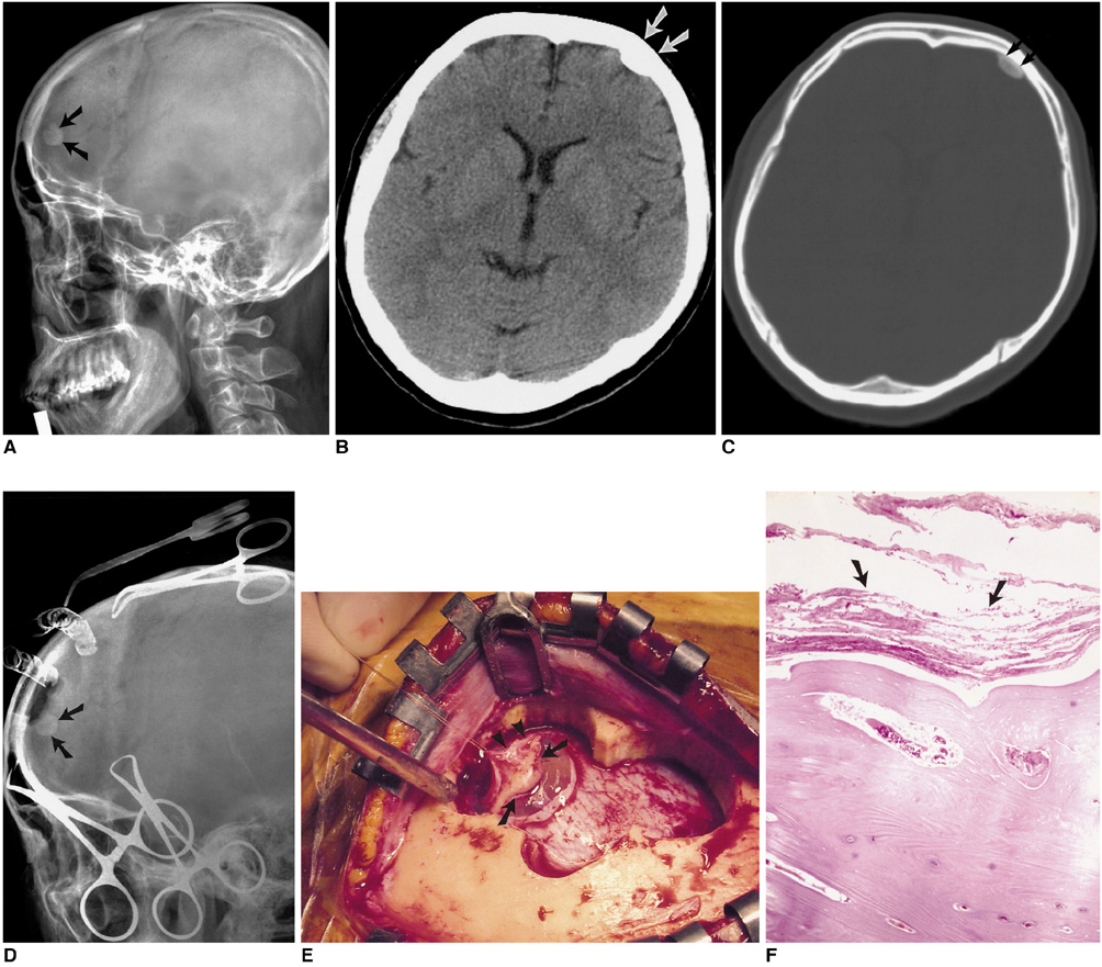
Fig. 1 A 43-year-old woman with subdural osteoma. A. Radiograph of the left lateral skull depicts a radiopaque lesion (arrows) in the frontal area. B. CT scan reveals the presence of a lentiform ossified lesion (arrows) in the left frontal area (window width: 90; window level: 25). C. Bone window setting shows a lucent line (arrows) between the ossified lesion and the inner table of the skull (window width: 2000; window level: 200). D. Intraoperative radiograph depicts an ossified lesion (arrows) under the craniectomy site. E. Intraoperative photograph indicates that the lesion (arrows) is firmly attached to the reflected dura (arrowheads). F. Microscopically, the tumor consists of a well-formed mature lamellar bone beneath the fibrous membrane (dura mater) (arrows) (H & E staining, ×40).
Cited by 1 articles
-
Clinical, Radiologic, and Pathologic Findings of Subdural Osteoma: A Case Report
Eun Young Kim, Yu Shik Shim, Dong Keun Hyun, Hyeonseon Park, Se Yang Oh, Seung Hwan Yoon
Brain Tumor Res Treat. 2016;4(1):40-43. doi: 10.14791/btrt.2016.4.1.40.
Reference
-
1. Fallon MD, Ellerbrake D, Teitelbaum SL. Meningeal osteomas and chronic renal failure. Hum Pathol. 1982. 13(5):449–453.2. Choudhury AR, Haleem A, Tjan GT. Solitary intradural intracranial osteoma. Br J Neurosurg. 1995. 9:557–559.3. Aoki H, Nakase H, Sakaki T. Subdural osteoma. Acta Neurochir (Wien). 1998. 140:727–728.4. Lee ST, Lui TN. Intracerebral osteoma: case report. Br J Neurosurg. 1997. 11:250–252.5. Vakaet A, De Reuck J, Thiery E, van der Eecken H. Intracerebral osteoma: a clinicopathologic and neuropsychologic case study. Child's Brain. 1983. 10:281–285.6. Avrahami E, Even I. Osteoma of the inner table of the skull-CT diagnosis. Clin Radiol. 2000. 55:435–438.
