Ten-year follow-up of full mouth rehabilitation with fixed prostheses using implants and natural tooth
- Affiliations
-
- 1Department of Prosthodontics, School of Dentistry, Jeonbuk National University, Jeonju, Republic of Korea
- 2Department of Dentistry, School of Medicine, Eulji University, Daejeon, Republic of Korea
- KMID: 2562763
- DOI: http://doi.org/10.14368/jdras.2024.40.3.189
Abstract
- In patients with newly established ideal occlusion through full-mouth rehabilitation using fixed prostheses, complications and occlusal changes over time can arise because of various factors. This case report describes a 10-year follow-up of a patient with masticatory dysfunction and aesthetic problems who underwent full-mouth rehabilitation with an increased vertical dimension. During the follow-up, complications such as tooth fracture, occlusal changes, infraocclusion of few implant-supported prostheses, and loss of interproximal contacts were observed. Detecting these issues early through periodic follow-up is important. This case report aims to review the causes of complications after full-mouth rehabilitation using fixed prostheses and the strategies for their management.
Figure
-
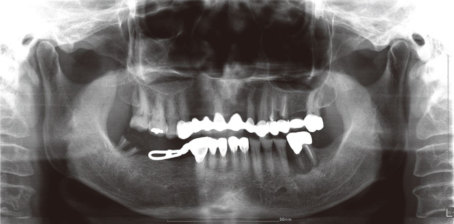
Fig. 1 Pre-treatment panoramic radiograph of the patient.
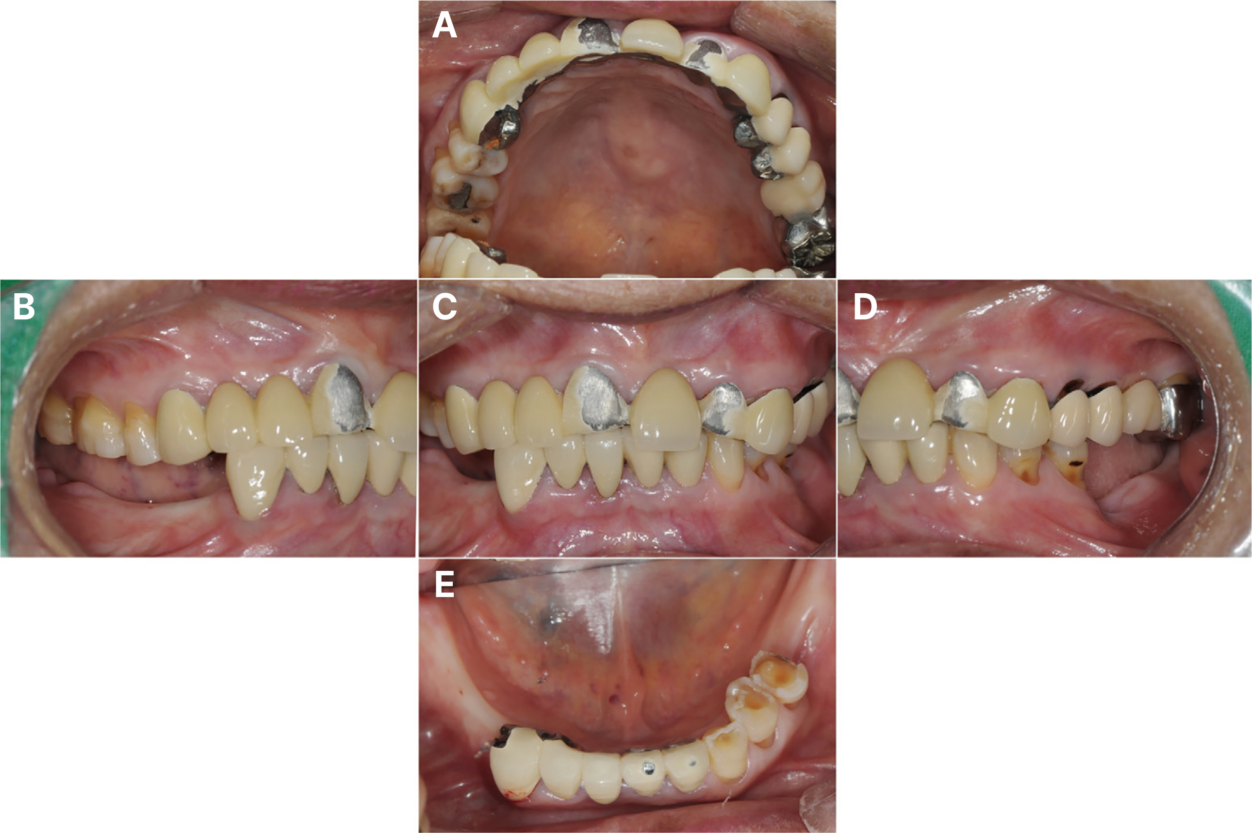
Fig. 2 Intra-oral status in the initial examination. (A) Maxillary occlusal view, (B) Right lateral view, (C) Frontal view at maximum inter-cuspal position, (D) Left lateral view, (E) Mandibular occlusal view.
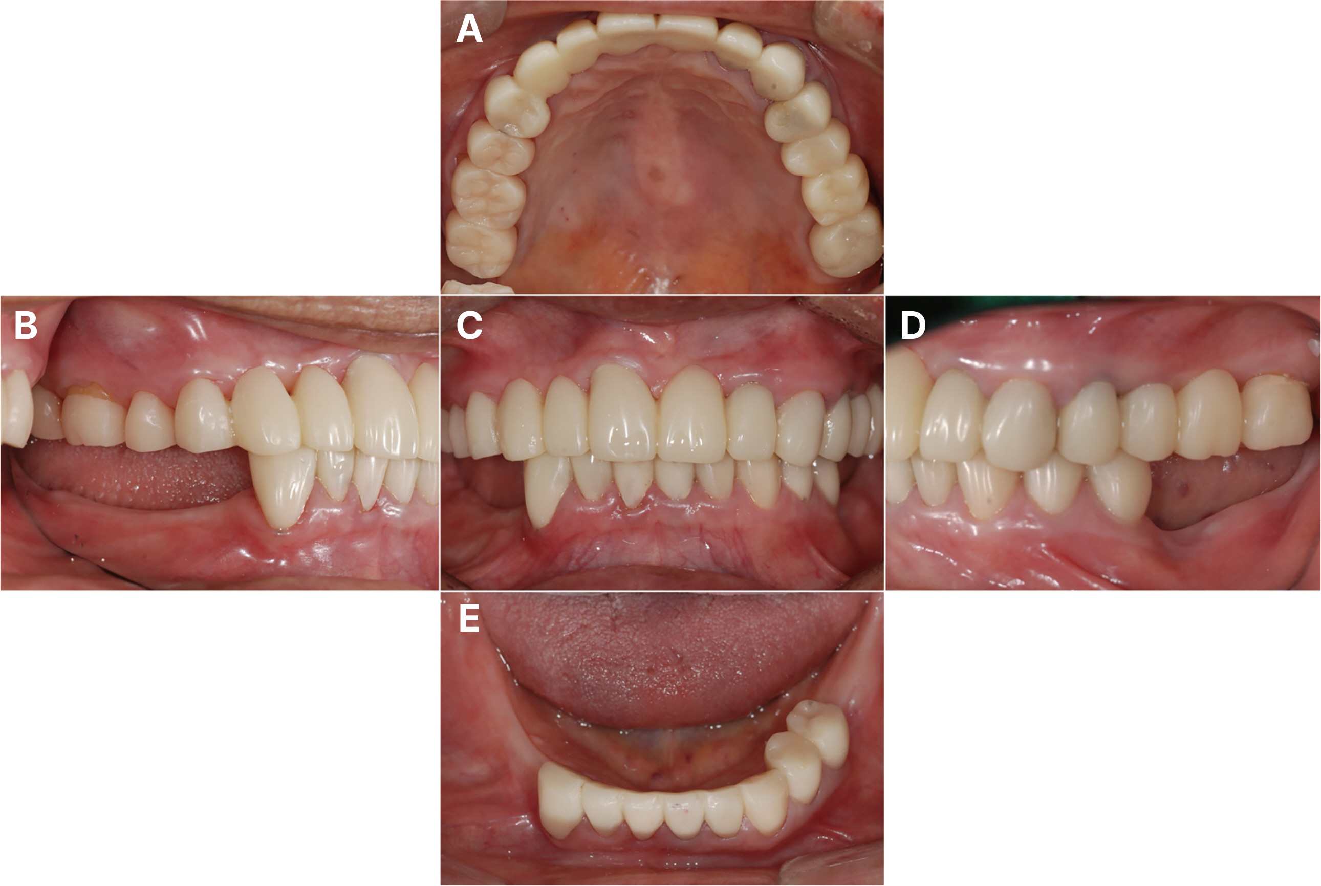
Fig. 3 1st provisional restoration. (A) Maxillary occlusal view, (B) Right lateral view, (C) Frontal view, (D) Left lateral view, (E) Mandibular occlusal view.

Fig. 4 Implant 1st surgery and GBR on #12, 13. (A) Creation of multiple perforations in the cortical bone of the recipient site with a round bur, (B) Bone graft with autogenous bone and xenogeneic bone (Geistlich Bio-Oss®), (C) Application of a resorbable collagen membrane (Geistlich Bio-Gide®), (D) Suture with 4-0 ethilon silk.
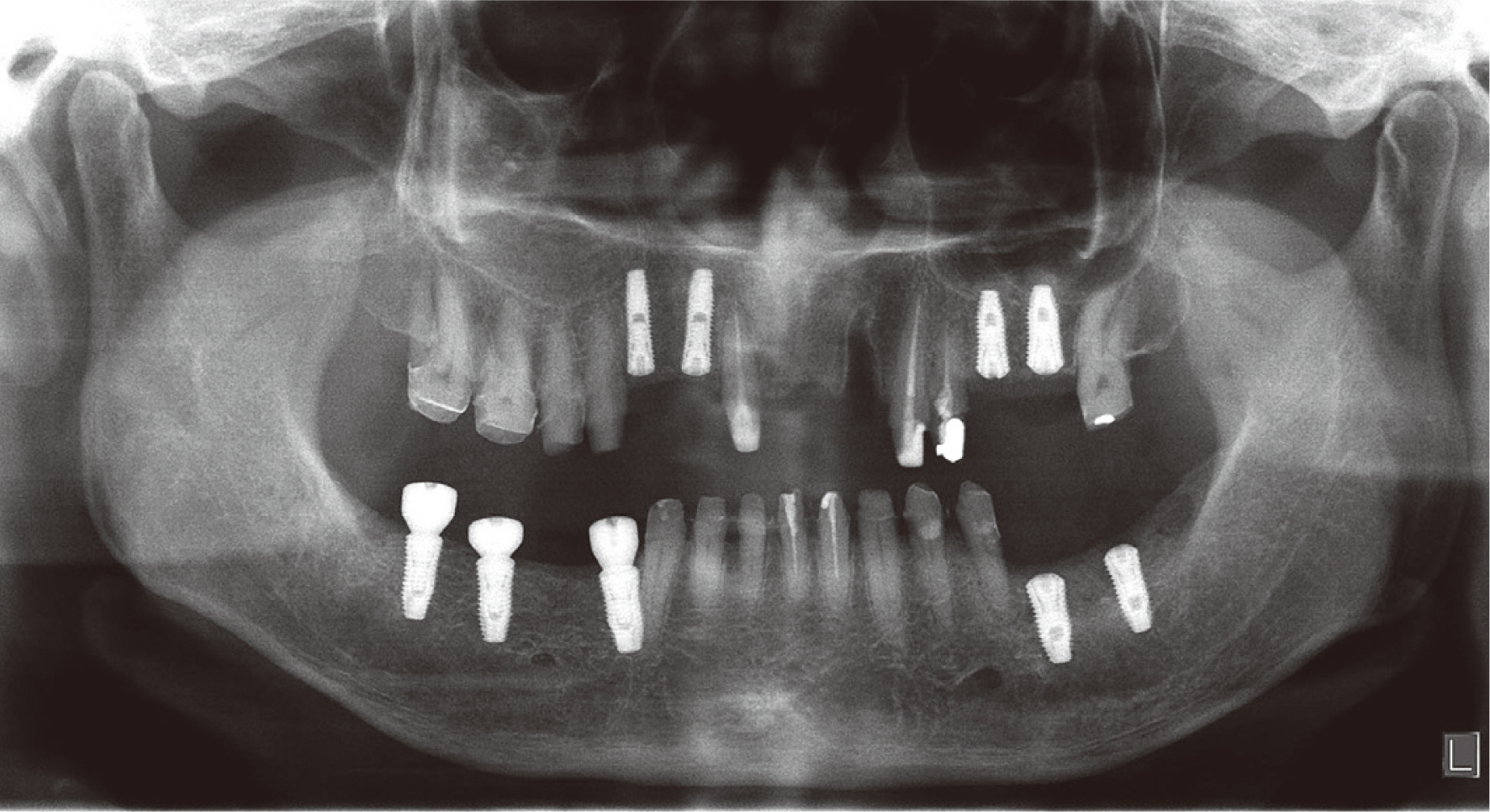
Fig. 5 Panoramic radiograph at implant placement.
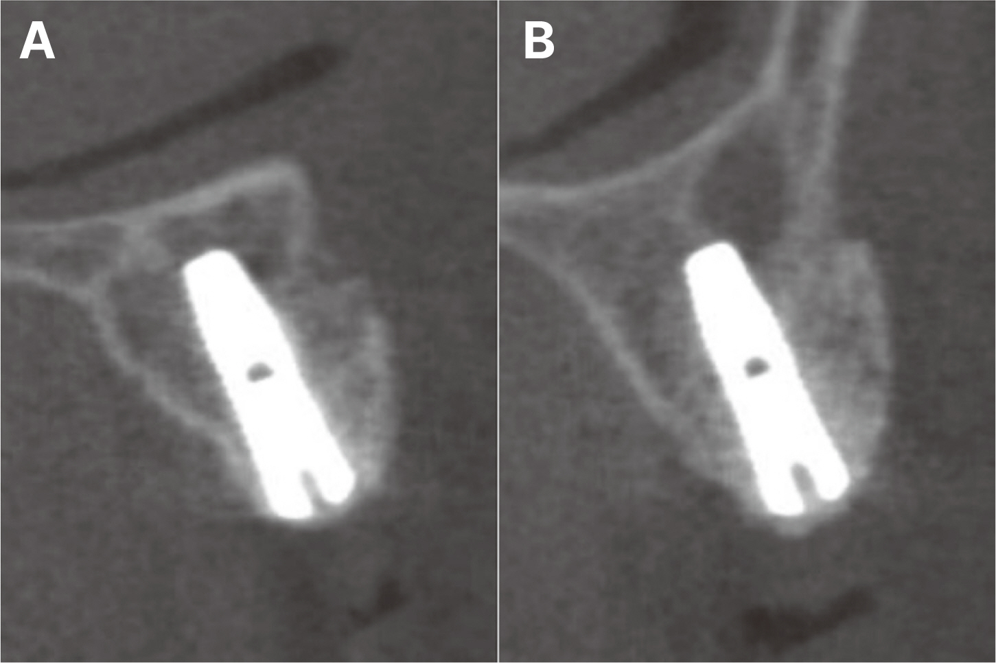
Fig. 6 6 months later CBCT radiograph after implantation and GBR. (A) #12i, (B) #13i.
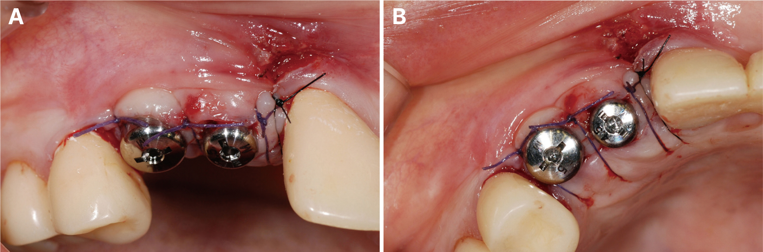
Fig. 7 2nd surgery on #12i, 13i. (A) Right lateral view, (B) Occlusal view.
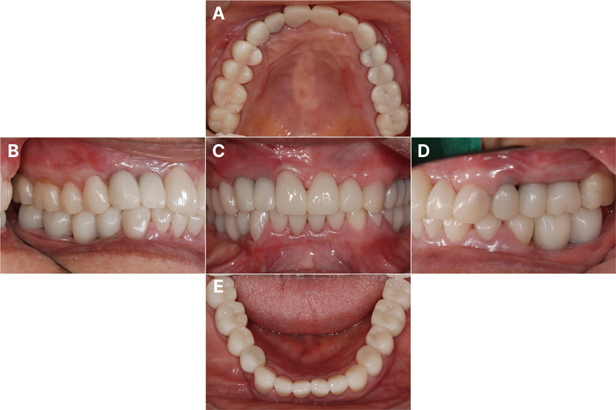
Fig. 8 2nd provisional restoration. (A) Maxillary occlusal view, (B) Right lateral view, (C) Frontal view, (D) Left lateral view, (E) Mandibular occlusal view.
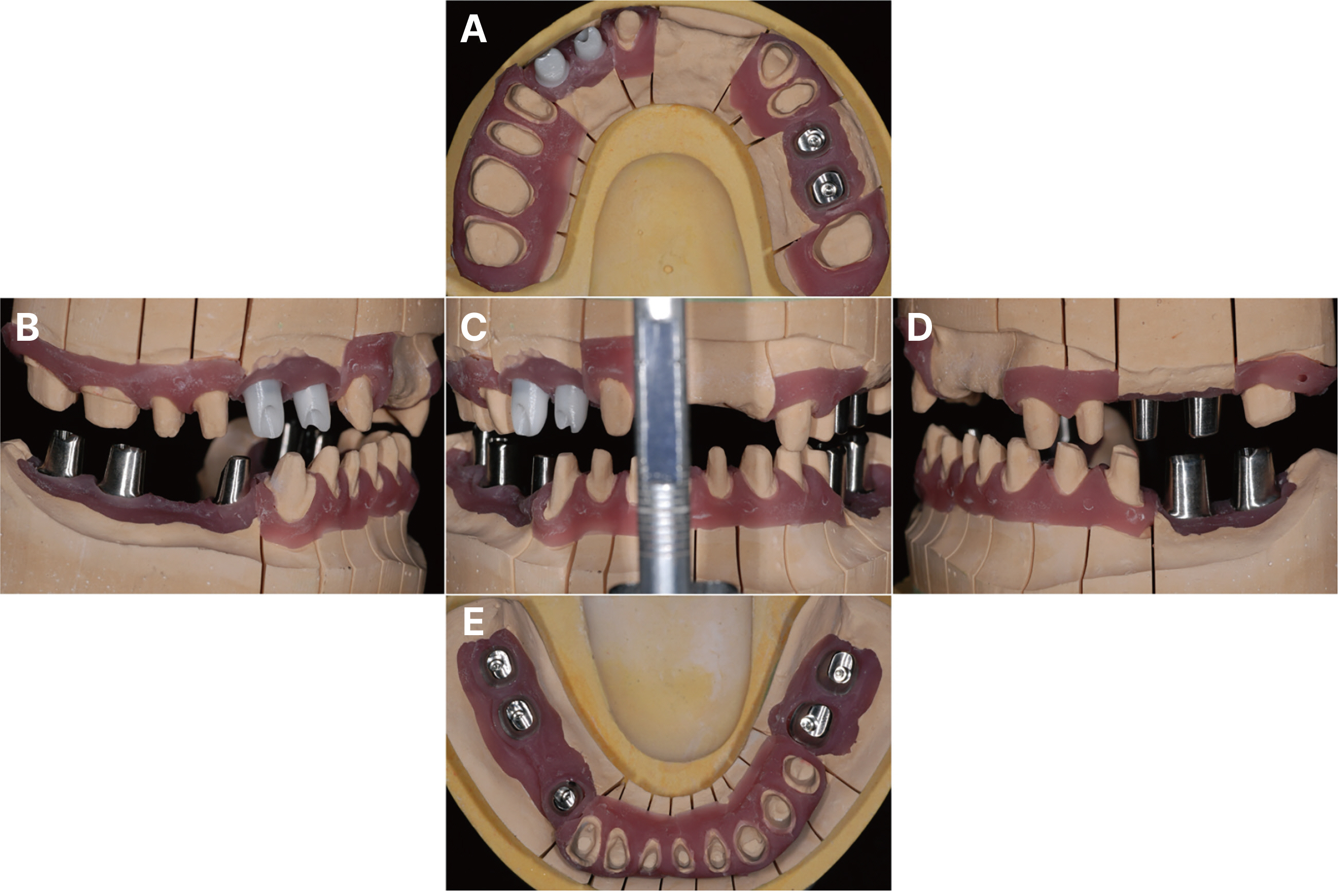
Fig. 9 Customized abutment for implant. (A) Maxillary occlusal view, (B) Right lateral view, (C) Frontal view, (D) Left lateral view, (E) Mandibular occlusal view.

Fig. 10 Definitive prosthesis was delivered. Esthetics and functions were restored with the zirconia prosthesis. (A) Maxillary occlusal view, (B) Right lateral view, (C) Frontal view at maximum inter-cuspal position, (D) Left lateral view, (E) Mandibular occlusal view.

Fig. 11 Panoramic radiograph when the definitive prosthesis delivered.

Fig. 12 Eccentric occlusion with definitive prosthesis. (A) Anterior movement: group function (guided by #11, 21), (B) Lateral movement - right side: group function (guided by #13i, 14), (C) Lateral movement - left side: canine guidance.
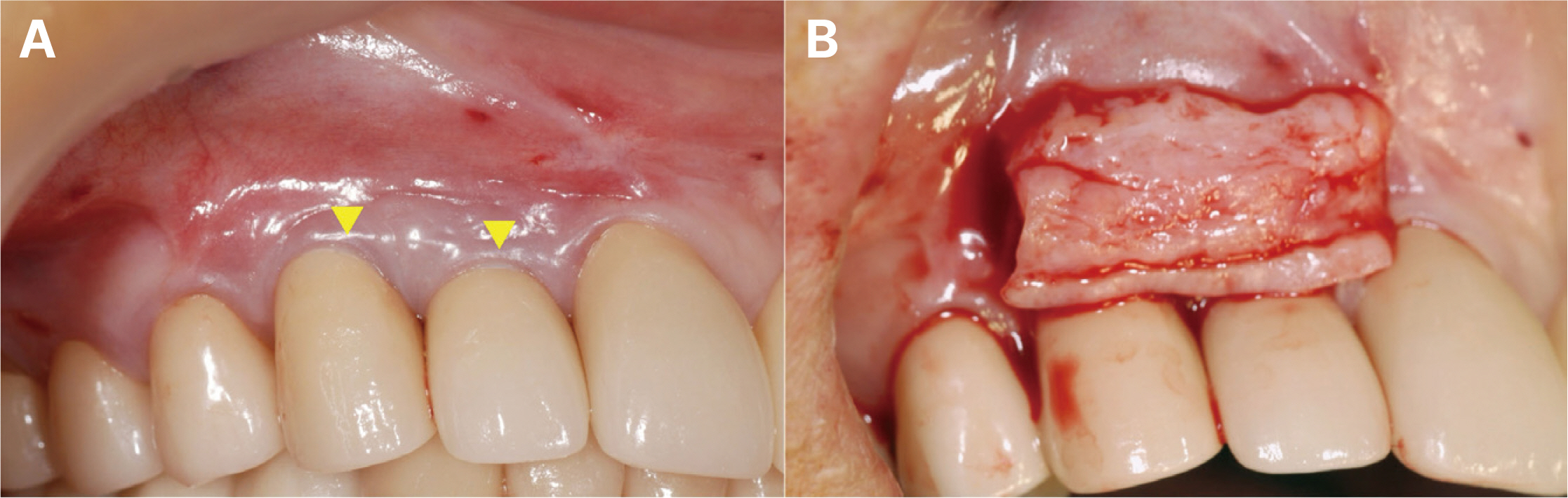
Fig. 13 Connective tissue graft on #12i, 13i. (A) Gingival recession defects on #12i, 13i, (B) Connective tissue graft for the treatment of the recession defects on #12i, 13i.
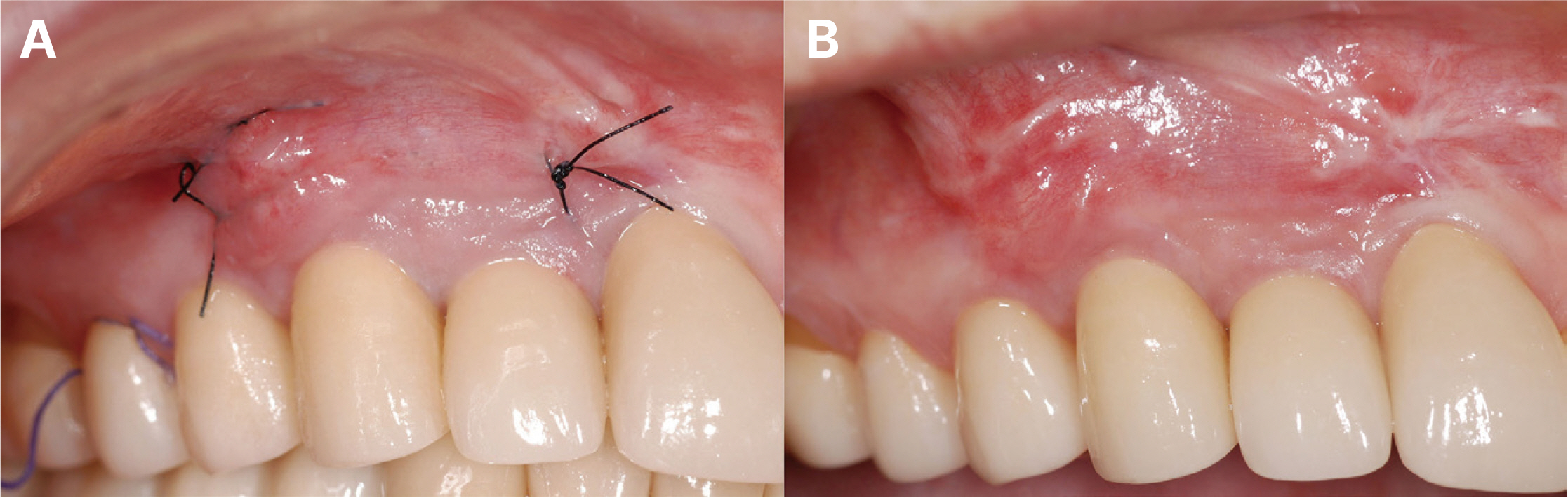
Fig. 14 Follow-up check on #12i, 13i connective tissue graft op site. (A) 1 week later check, (B) 4 weeks later check.

Fig. 15 Follow-up check after 6 years. (A) Occlusal view when the definitive prosthesis delivered, (B) Occlusal view after 6 years with #25i, 26i infraocclusion.
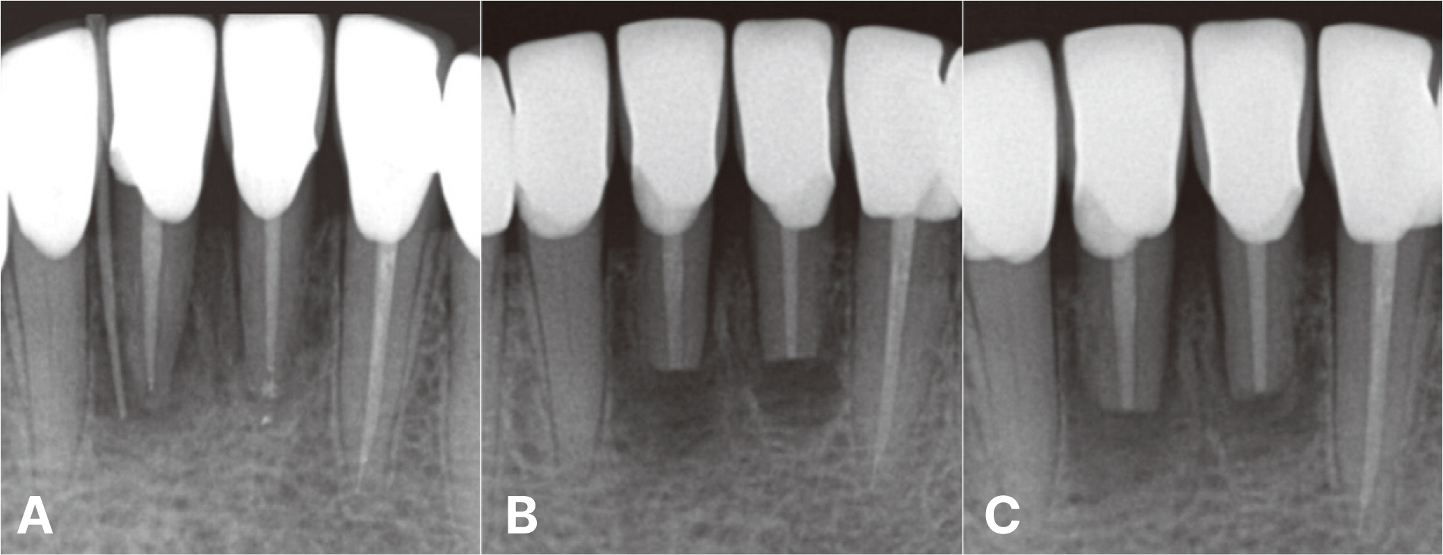
Fig. 16 Follow-up check after 6 years. (A) #31 mesial deep pocket depth, GP cone tracing on #41 labial fistula, (B) #31, 41 apicoectomy, (C) 1 month follow-up check after apicoectomy.

Fig. 17 Follow-up check after 7 years. #25i, 36i mesial interproximal contact loss.
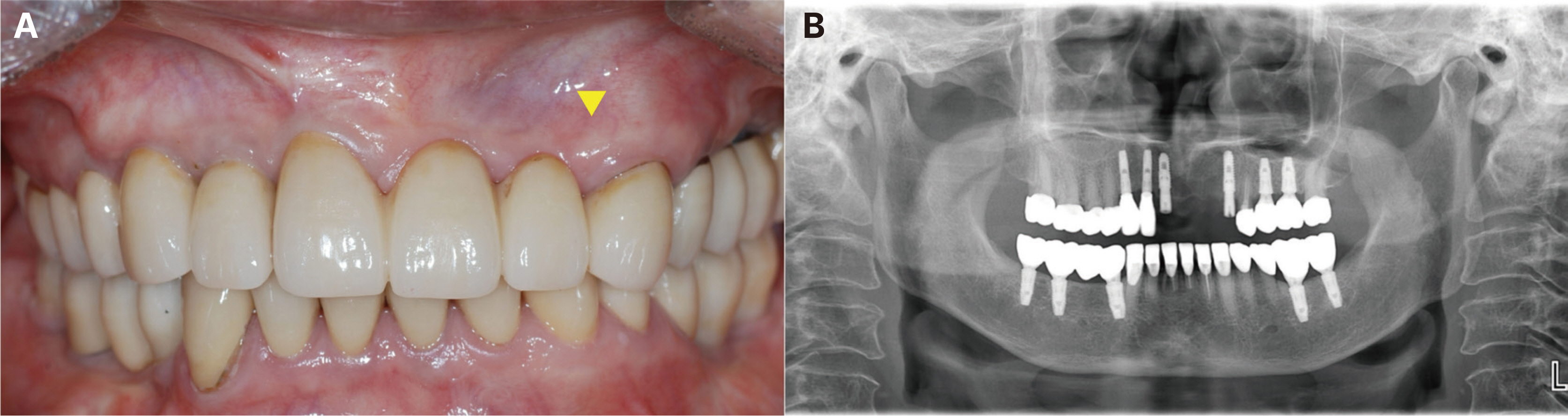
Fig. 18 Follow-up check after 7 years. (A) #23 gingival swelling, (B) #11, 23 immediate implant placement.
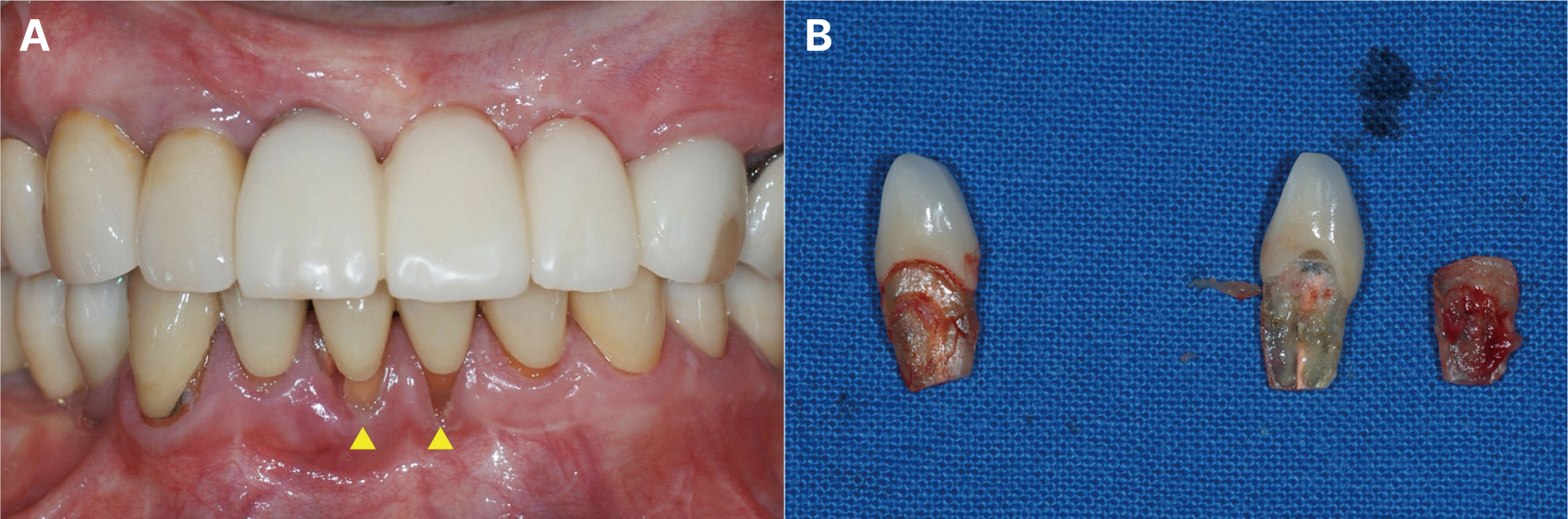
Fig. 19 Follow-up check after 8 years. (A) Poor prognosis of #31, 41, (B) Extraction of #31, 41.

Fig. 20 Follow-up check after 8 years. #11i-23i 4 unit bridge, #32-42 4 unit bridge were delivered.
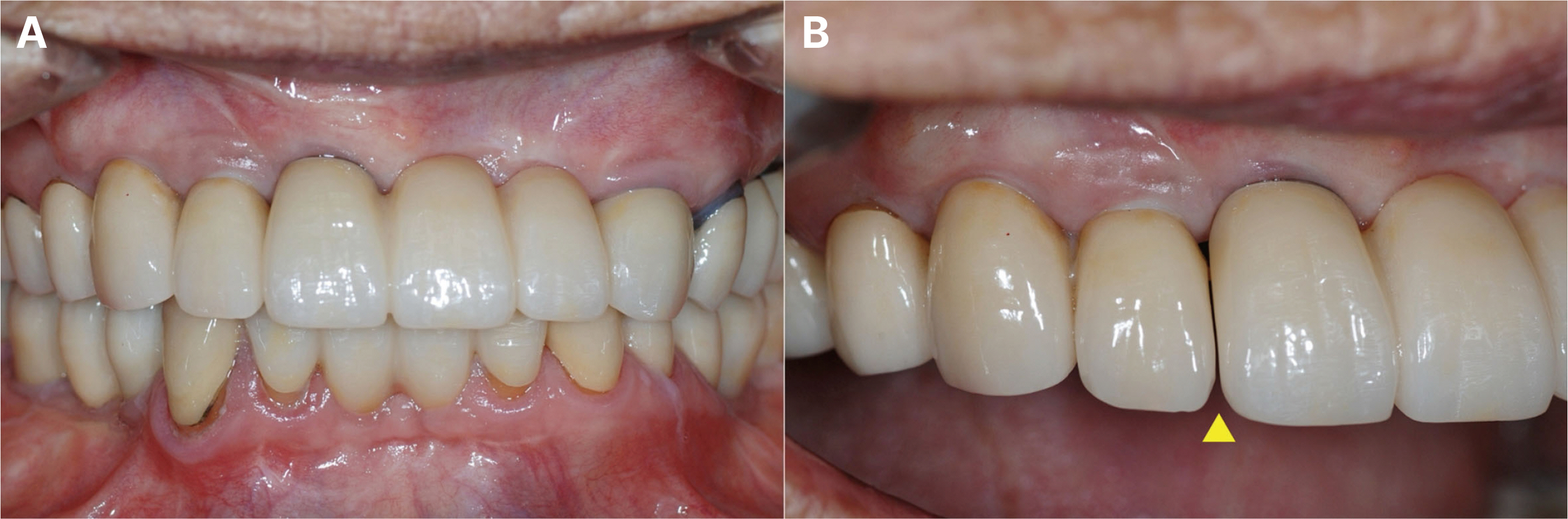
Fig. 21 Follow-up check after 9 years. #11i-12i interproximal contact loss. (A) Frontal view, (B) Right lateral view.
Reference
-
References
1. Wolpoff MH. 1971; Interstitial wear. Am J Phys Anthropol. 34:205–27. DOI: 10.1002/ajpa.1330340206. PMID: 5103127.2. Jo DW, Kwon MJ, Kim JH, Kim YK, Yi YJ. 2019; Evaluation of adjacent tooth displacement in the posterior implant restoration with proximal contact loss by superimposition of digital models. J Adv Prosthodont. 11:88–94. DOI: 10.4047/jap.2019.11.2.88. PMID: 31080569. PMCID: PMC6491362.3. Bishara SE, Treder JE, Jakobsen JR. 1994; Facial and dental changes in adulthood. Am J Orthod Dentofacial Orthop. 106:175–86. DOI: 10.1016/S0889-5406(94)70036-2. PMID: 8059754.4. Heij DG, Opdebeeck H, van Steenberghe D, Kokich VG, Belser U, Quirynen M. 2006; Facial development, continuous tooth eruption, and mesial drift as compromising factors for implant placement. Int J Oral Maxillofac Implants. 21:867–78. DOI: 10.1016/s0022-3913(07)60086-9. PMID: 17190296.5. Wei H, Tomotake Y, Nagao K, Ichikawa T. 2008; Implant prostheses and adjacent tooth migration: preliminary retrospective survey using 3-dimensional occlusal analysis. Int J Prosthodont. 21:302–4. PMID: 18717086.6. Daftary F, Mahallati R, Bahat O, Sullivan RM. 2013; Lifelong craniofacial growth and the implications for osseointegrated implants. Int J Oral Maxillofac Implants. 28:163–9. DOI: 10.11607/jomi.2827. PMID: 23377062.7. Park YH, Kim KA, Lee JJ, Seo JM. 2024; Interproximal contact loss between implant-supported prostheses: a clinical report. J Korean Acad Prosthodont. 62:47–53. DOI: 10.4047/jkap.2024.62.1.47.8. Byun SJ, Heo SM, Ahn SG, Chang M. 2015; Analysis of proximal contact loss between implant-supported fixed dental prostheses and adjacent teeth in relation to influential factors and effects. A cross-sectional study. Clin Oral Implants Res. 26:709–14. DOI: 10.1111/clr.12373. PMID: 24712313.9. Varthis S, Randi A, Tarnow DP. 2016; Prevalence of interproximal open contacts between single-implant restorations and adjacent teeth. Int J Oral Maxillofac Implants. 31:1089–92. DOI: 10.11607/jomi.4432. PMID: 27632264.10. French D, Naito M, Linke B. 2019; Interproximal contact loss in a retrospective cross-sectional study of 4325 implants: Distribution and incidence and the effect on bone loss and peri-implant soft tissue. J Prosthet Dent. 122:108–14. DOI: 10.1016/j.prosdent.2018.11.011. PMID: 30885585.11. Lee JH, Kim DG, Park CJ, Cho LR. 2014; Axial displacements in external and internal implant-abutment connection. Clin Oral Implants Res. 25:e83–9. DOI: 10.1111/clr.12062. PMID: 23088616.12. Jemt T. 2005; Measurements of tooth movements in relation to single-implant restorations during 16 years: a case report. Clin Implant Dent Relat Res. 7:200–8. DOI: 10.1111/j.1708-8208.2005.tb00065.x. PMID: 16336911.13. Schwartz NL, Whitsett LD. 1970; Berry TG, Stewart JL. Unserviceable crowns and fixed partial dentures: life-span and causes for loss of serviceability. J Am Dent Assoc. 81:395–401. DOI: 10.14219/jada.archive.1970.0398. PMID: 5273607.14. Libby G, Arcuri MR, LaVelle WE, Hebl L. 1997; Longevity of fixed partial dentures. J Prosthet Dent. 78:127–31. DOI: 10.1016/S0022-3913(97)70115-X. PMID: 9260128.15. Lang NP, Zitzmann NU. 2012; Clinical research in implant dentistry: evaluation of implant-supported restorations, aesthetic and patient-reported outcomes. J Clin Periodontol. 39 Suppl 12:133–8. DOI: 10.1111/j.1600-051X.2011.01842.x. PMID: 22533953.16. Turner KA, Missirlian DM. 1984; Restoration of the extremely worn dentition. J Prosthet Dent. 52:467–74. DOI: 10.1016/0022-3913(84)90326-3. PMID: 6389829.17. Wood GN. 1988; Centric relation and the treatment position in rehabilitating occlusions: a physiologic approach. Part II: The treatment position. J Prosthet Dent. 60:15–8. DOI: 10.1016/0022-3913(88)90341-1. PMID: 3165459.18. Sfondouris T, Prestipino V. 2019; Chairside management of an open proximal contact on an implant-supported ceramic crown using direct composite resin. J Prosthet Dent. 122:1–4. DOI: 10.1016/j.prosdent.2018.10.019. PMID: 30948300.19. Landys Borén D, Jonasson P, Kvist T. 2015; Long-term survival of endodontically treated teeth at a public dental specialist clinic. J Endod. 41:176–81. DOI: 10.1016/j.joen.2014.10.002. PMID: 25453569.20. Burke FJT, Lucarotti PSK, Wilson N. 2021; Canine guidance on crowned teeth: time for a rethink? Br Dent J. 230:285–8. DOI: 10.1038/s41415-021-2699-3. PMID: 33712777.
- Full Text Links
-
- Actions
-
Cited
- CITED
-
- Close
- Share
-
- Similar articles
-
- Full mouth fixed implant rehabilitation in a patient with generalized aggressive periodontitis
- Full-mouth rehabilitation with implant-supported fixed dental prostheses for the edentulous maxilla and partially edentulous mandible: A case report
- Three-year follow-up of full mouth rehabilitation with anterior implant surveyed bridges and distal extension removable partial denture
- Full mouth rehabilitation of edentulous patient with intellectual disability using implants and monolithic zirconia
- A stress analysis of fixed prostheses with dental implant and natural tooth
