Ann Lab Med.
2024 Nov;44(6):572-575. 10.3343/alm.2024.0003.
Reliability of the DI-60 Digital Image Analyzer for Detecting Platelet Clumping and Obtaining Accurate Platelet Counts
- Affiliations
-
- 1Department of Laboratory Medicine, Soonchunhyang University Cheonan Hospital, Soonchunhyang University College of Medicine, Cheonan, Korea
- 2Clinical Trial Center, Soonchunhyang University Cheonan Hospital, Soonchunhyang University College of Medicine, Cheonan, Korea
- KMID: 2560804
- DOI: http://doi.org/10.3343/alm.2024.0003
Abstract
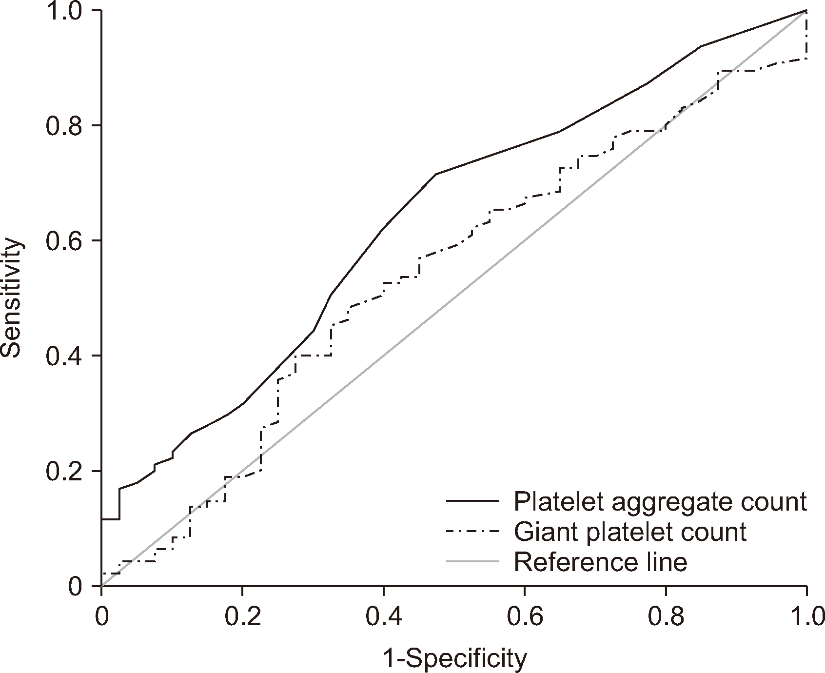
- Pseudothrombocytopenia caused by platelet clumping (PC) can lead to unnecessary platelet transfusions or underdiagnosis of hematologic neoplasms. To overcome these limitations, we assessed the capacity of the Sysmex DI-60 digital morphology analyzer (Sysmex, Kobe, Japan) for detecting PC and determining an accurate platelet count in the presence of PC. For this purpose, 135 samples with or without PC (groups Y and N, respectively) were processed by an examiner (a hematologic specialist) using both the Sysmex XN-9000 and DI-60 analyzers. Although the platelet aggregate (PA) and giant platelet (GP) counts reported by the DI-60 and the examiner exhibited strong correlations, they proved inadequate as effective indicators for screening samples containing PC. Between the PA and GP counts and four platelet indices (the platelet distribution width [PDW], mean platelet volume [MPV], platelet large cell ratio [P_LCR], and plateletcrit [PCT]) reported by the XN-9000, we observed statistically significant correlations (both overall and with group Y), but they were relatively weak. The platelet counts determined using the DI-60 and light microscopy in group Y showed substantial variations. Although the performance of the DI-60 was reliable for detecting PA and GP in smear images, such fixed areas are not representative of whole samples. Further, in the presence of PC, the resulting platelet counts determined using the DI-60 were not sufficiently accurate to be accepted as the final count.
Keyword
Figure
Reference
-
References
1. Lardinois B, Favresse J, Chatelain B, Lippi G, Mullier F. 2021; Pseudothrombocytopenia-a review on causes, occurrence and clinical implications. J Clin Med. 10:594. DOI: 10.3390/jcm10040594. PMID: 33557431. PMCID: PMC7915523. PMID: d53aef395a6d4be6951f486001d28eb1.2. Baccini V, Geneviève F, Jacqmin H, Chatelain B, Girard S, Wuilleme S, et al. 2020; Platelet counting: ugly traps and good advice. Proposals from the French-speaking Cellular Hematology Group (GFHC). J Clin Med. 9:808. DOI: 10.3390/jcm9030808. PMID: 32188124. PMCID: PMC7141345. PMID: 704f5f47e8944dcf89f9bf3558e2f964.3. Bizzaro N. 1995; EDTA-dependent pseudothrombocytopenia: a clinical and epidemiological study of 112 cases, with 10-year follow-up. Am J Hematol. 50:103–9. DOI: 10.1002/ajh.2830500206. PMID: 7572988.4. Choe WH, Cho YU, Chae JD, Kim SH. 2013; Pseudothrombocytopenia or platelet clumping as a possible cause of low platelet count in patients with viral infection: a case series from single institution focusing on hepatitis A virus infection. Int J Lab Hematol. 35:70–6. DOI: 10.1111/j.1751-553X.2012.01466.x. PMID: 22958573.5. Kratz A, Lee SH, Zini G, Riedl JA, Hur M, Machin S, et al. 2019; Digital morphology analyzers in hematology: ICSH review and recommendations. Int J Lab Hematol. 41:437–47. DOI: 10.1111/ijlh.13042. PMID: 31046197.6. Billard M, Lainey E, Armoogum P, Alberti C, Fenneteau O, Da Costa L. 2010; Evaluation of the CellaVision DM automated microscope in pediatrics. Int J Lab Hematol. 32:530–8. DOI: 10.1111/j.1751-553X.2009.01219.x. PMID: 20132350.7. Gulati G, Uppal G, Florea AD, Gong J. 2014; Detection of platelet clumps on peripheral blood smears by CellaVision DM96 system and microscopic review. Lab Med. 45:368–71. DOI: 10.1309/LM604RQVKVLRFXOR. PMID: 25316670.8. Gao Y, Mansoor A, Wood B, Nelson H, Higa D, Naugler C. 2013; Platelet count estimation using the CellaVision DM96 system. J Pathol Inform. 4:16. DOI: 10.4103/2153-3539.114207. PMID: 23858391. PMCID: PMC3709429. PMID: d0df0893d0284640bc3763bf02e0f263.9. Gulati G, Caro J. 2014. Blood cells: morphology and clinical relevance. 2nd ed. American Society for Clinical Pathology;Chicago: p. 94.10. Mannuß S. 2020; Influence of different methods and anticoagulants on platelet parameter measurement. J Lab Med. 44:255–72. DOI: 10.1515/labmed-2020-0037. PMID: d188b7b31a0844bb8b0c934e06005564.11. Nam M, Yoon S, Hur M, Lee GH, Kim H, Park M, et al. 2022; Digital Morphology Analyzer Sysmex DI-60 vs. Manual Counting for White Blood Cell Differentials in Leukopenic Samples: A Comparative Assessment of Risk and Turnaround Time. Ann Lab Med. 42:398–405. DOI: 10.3343/alm.2022.42.4.398. PMID: 35177560. PMCID: PMC8859564.12. Kim HN, Hur M, Kim H, Kim SW, Moon HW, Yun YM. 2017; Performance of automated digital cell imaging analyzer Sysmex DI-60. Clin Chem Lab Med. 56:94–102. DOI: 10.1515/cclm-2017-0132. PMID: 28672770.13. Briggs C, Harrison P, Machin SJ. 2007; Continuing developments with the automated platelet count. Int J Lab Hematol. 29:77–91. DOI: 10.1111/j.1751-553X.2007.00909.x. PMID: 17474881.14. ICSH. 2001; Platelet counting by the RBC/platelet ratio method: A reference method. Am J Clin Pathol. 115:460–4. DOI: 10.1309/W612-MYEP-FA7U-8UYA. PMID: 11246942.
- Full Text Links
- Actions
-
Cited
- CITED
-
- Close
- Share
- Similar articles
-
- Integration of an MC-80 Digital Image Analyzer With an Automated BC-6800Plus Hematology Analyzer Enables Accurate Platelet Counting in Samples With EDTA-Induced Pseudothrombocytopenia
- Correction of Platelet Count Using a Vortex in Pseudothrombocytopenia
- Pseudothrombocytopenia Due to Anticoagulant-Independent Agglutinins
- Evaluation of Platelet count by the CELL-DYN Sapphire CD61 Immunoplatelet Method in Patients with Hematologic Diseases Receiving Chemotherapy
- Bleeding time: Remained or Retired or Removed?


