Ann Rehabil Med.
2024 Oct;48(5):352-359. 10.5535/arm.240041.
Change in Plantar Pressure and Plain Radiography in Pediatric Flexible Flatfoot: A Retrospective Cohort Study
- Affiliations
-
- 1Department of Rehabilitation Medicine, Hanyang University Medical Center, Hanyang University College of Medicine, Seoul, Korea
- KMID: 2560473
- DOI: http://doi.org/10.5535/arm.240041
Abstract
Objective
To investigate longitudinal changes in plantar pressure examinations and plain radiographs and to assess the correlations between these two modalities in pediatric patients with flexible flatfoot (FFF).
Methods
Pediatric patients diagnosed with FFF were analyzed in this retrospective cohort study. Medical records were reviewed to collect data on plain radiographs and plantar pressure examinations. Changes in radiographic angles and plantar pressure parameters were analyzed over a follow-up period exceeding 1 year. Statistical analyses included paired t-test, Wilcoxon signed rank test, and Spearman’s correlation analysis.
Results
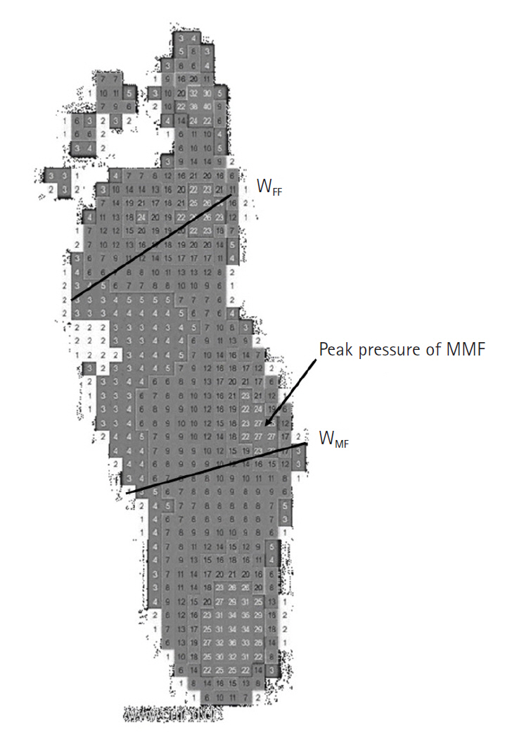
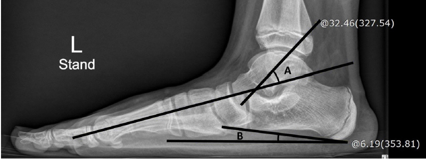
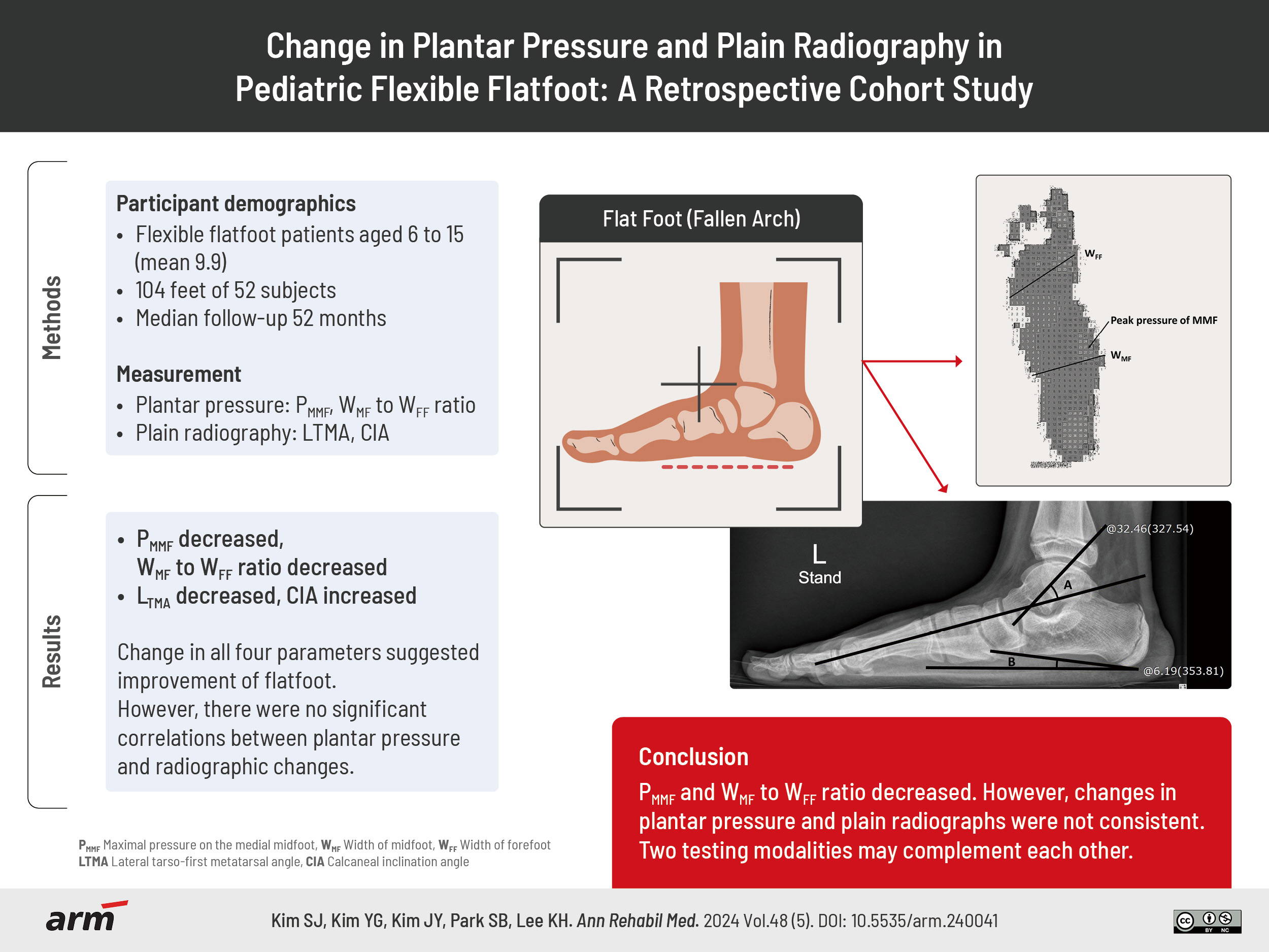
A total of 52 subjects was included in the plantar pressure analysis, with a mean age of 9.9 years at the first visit and a median follow-up duration of 52 months. The lateral tarso-first metatarsal angle decreased by 1.3° (p<0.001) and calcaneal inclination angle increased by 2.5° (p<0.001) in these patients. The percentage value of midfoot width (WMF) divided by forefoot width (WFF) decreased by an average of 9.8% p (p<0.001), and the maximal pressure on the medial midfoot (MMF) decreased by 1.6 N/cm2 (p<0.001). However, no correlations were found between plantar pressure and radiographic changes.
Conclusion
During follow-up of patients with FFF, the maximal pressure on the MMF and the ratio of WMF to WFF decreased in successive plantar pressure examinations. Changes observed in plantar pressure and plain radiographs were not consistent, indicating that these two testing modalities can complement each other.
Keyword
Figure
Reference
-
1. Pfeiffer M, Kotz R, Ledl T, Hauser G, Sluga M. Prevalence of flat foot in preschool-aged children. Pediatrics. 2006; 118:634–9.
Article2. Rodriguez N, Volpe RG. Clinical diagnosis and assessment of the pediatric pes planovalgus deformity. Clin Podiatr Med Surg. 2010; 27:43–58.
Article3. Park GY, Lee WC. Prevalence of the flatfoot and its relation with the practice of wearing footwear of primary school children in Korea. J Korean Acad Rehabil Med. 2001; 25:867–76.4. Harris EJ. The natural history and pathophysiology of flexible flatfoot. Clin Podiatr Med Surg. 2010; 27:1–23.
Article5. Harris EJ, Vanore JV, Thomas JL, Kravitz SR, Mendelson SA, Mendicino RW, Clinical Practice Guideline Pediatric Flatfoot Panel of the American College of Foot and Ankle Surgeons, et al. Diagnosis and treatment of pediatric flatfoot. J Foot Ankle Surg. 2004; 43:341–73.
Article6. Choi JY, Lee DJ, Kim SJ, Suh JS. Does the long-term use of medial arch support insole induce the radiographic structural changes for pediatric flexible flat foot? - a prospective comparative study. Foot Ankle Surg. 2020; 26:449–56.
Article7. Kanatlı U, Aktas E, Yetkin H. Do corrective shoes improve the development of the medial longitudinal arch in children with flexible flat feet? J Orthop Sci. 2016; 21:662–6.
Article8. Cho DJ, Ahn SY, Bok SK. Effect of foot orthoses in children with symptomatic flexible flatfoot based on ultrasonography of the ankle invertor and evertor muscles. Ann Rehabil Med. 2021; 45:459–70.
Article9. Chen KC, Chen YC, Yeh CJ, Hsieh CL, Wang CH. The effect of insoles on symptomatic flatfoot in preschool-aged children: a prospective 1-year follow-up study. Medicine (Baltimore). 2019; 98:e17074.10. Bok SK, Kim BO, Lim JH, Ahn SY. Effects of custom-made rigid foot orthosis on pes planus in children over 6 years old. Ann Rehabil Med. 2014; 38:369–75.
Article11. Youn KJ, Ahn SY, Kim BO, Park IS, Bok SK. Long-term effect of rigid foot orthosis in children older than six years with flexible flat foot. Ann Rehabil Med. 2019; 43:224–9.
Article12. Evans AM, Rome K, Carroll M, Hawke F. Foot orthoses for treating paediatric flat feet. Cochrane Database Syst Rev. 2022; 1:CD006311.
Article13. Lee HJ, Lim KB, Yoo J, Yoon SW, Yun HJ, Jeong TH. Effect of custom-molded foot orthoses on foot pain and balance in children with symptomatic flexible flat feet. Ann Rehabil Med. 2015; 39:905–13.
Article14. Shin BJ, Lee KM, Chung CY, Sung KH, Chun DI, Hong CH, et al. Analysis of factors influencing improvement of idiopathic flatfoot. Medicine (Baltimore). 2021; 100:e26894.
Article15. Park MS, Kwon SS, Lee SY, Lee KM, Kim TG, Chung CY. Spontaneous improvement of radiographic indices for idiopathic planovalgus with age. J Bone Joint Surg Am. 2013; 95:e193(1-8).
Article16. Seol YJ, Jung ST, Yang HK, Lee KB, Oh CS, Jung YJ, et al. Diagnostic availability of pedobarography and correlation of radiographic and pedobarographic measurements in pediatric flexible flatfoot. J Korean Orthop Assoc. 2014; 49:366–73.
Article17. Kadhim M, Holmes L Jr, Miller F. Correlation of radiographic and pedobarograph measurements in planovalgus foot deformity. Gait Posture. 2012; 36:177–81.
Article18. Hennig EM, Staats A, Rosenbaum D. Plantar pressure distribution patterns of young school children in comparison to adults. Foot Ankle Int. 1994; 15:35–40.
Article19. Onodera AN, Sacco IC, Morioka EH, Souza PS, de Sá MR, Amadio AC. What is the best method for child longitudinal plantar arch assessment and when does arch maturation occur? Foot (Edinb). 2008; 18:142–9.
Article20. Miller CA, Verstraete MC. Determination of the step duration of gait initiation using a mechanical energy analysis. J Biomech. 1996; 29:1195–9.
Article21. Yildiz K, Cetin T. Interobserver reliability in the radiological evaluation of flatfoot (pes planus) deformity: a cross-sectional study. J Foot Ankle Surg. 2022; 61:1065–70.
Article22. Younger AS, Sawatzky B, Dryden P. Radiographic assessment of adult flatfoot. Foot Ankle Int. 2005; 26:820–5.
Article23. Bock P, Pittermann M, Chraim M, Rois S. The inter- and intraobserver reliability for the radiological parameters of flatfoot, before and after surgery. Bone Joint J. 2018; 100-B:596–602.
Article24. Chen KC, Tung LC, Yeh CJ, Yang JF, Kuo JF, Wang CH. Change in flatfoot of preschool-aged children: a 1-year follow-up study. Eur J Pediatr. 2013; 172:255–60.
Article25. Lee JS, Kim KB, Jeong JO, Kwon NY, Jeong SM. Correlation of foot posture index with plantar pressure and radiographic measurements in pediatric flatfoot. Ann Rehabil Med. 2015; 39:10–7.
Article26. Lee SW, Choi JH, Kwon HJ, Song KS. Effect of pressure based customized 3-dimensional printing insole in pediatric flexible flat foot patients. J Korean Foot Ankle Soc. 2020; 24:113–9.
Article27. Paecharoen S, Arunakul M, Tantivangphaisal N. Diagnostic accuracy of harris imprint index, chippaux-smirak index, staheli index compared with talar-first metatarsal angle for screening arch of foot. Ann Rehabil Med. 2023; 47:222–7.
Article28. Pehlivan O, Cilli F, Mahirogullari M, Karabudak O, Koksal O. Radiographic correlation of symptomatic and asymptomatic flexible flatfoot in young male adults. Int Orthop. 2009; 33:447–50.
Article29. Moraleda L, Mubarak SJ. Flexible flatfoot: differences in the relative alignment of each segment of the foot between symptomatic and asymptomatic patients. J Pediatr Orthop. 2011; 31:421–8.30. Lee YK, Yim SJ, Lee SH, Park CH, Lee SH. The talus-1st metatarsal angle, the talo-horizontal angle and calcaneal pitch angle of young men in Korea. J Korean Foot Ankle Surg. 2010; 14:161–4.
- Full Text Links
- Actions
-
Cited
- CITED
-
- Close
- Share
- Similar articles
-
- Correlation of Foot Posture Index With Plantar Pressure and Radiographic Measurements in Pediatric Flatfoot
- Diagnostic Availability of Pedobarography and Correlation of Radiographic and Pedobarographic Measurements in Pediatric Flexible Flatfoot
- Operative Treatment of Adult Flexible Flatfoot with Young's Tenosuspension: Case Report
- Effect of Pressure Based Customized 3-Dimensional Printing Insole in Pediatric Flexible Flat Foot Patients
- Adult Idiopathic Flexible Flat Foot Treated with Medial Sliding Calcaneal Osteotomy and Subtalar Arthroereisis: Report of 1 Case


