Effect of Pressure Based Customized 3-Dimensional Printing Insole in Pediatric Flexible Flat Foot Patients
- Affiliations
-
- 1Department of Orthopedic Surgery, Keimyung University School of Medicine, Daegu, Korea
- KMID: 2506528
- DOI: http://doi.org/10.14193/jkfas.2020.24.3.113
Abstract
- Purpose
A flatfoot that fails to form a longitudinal foot arch is a common lower limb deformity in children. This study evaluated the structural and functional effects of the insole for pediatric flexible flat foot (PFFF).
Materials and Methods
Twenty-nine PFFF patients (20 boys and 9 girls, 58 feet) with bilateral symptomatic flatfoot deformities between February 2017 and May 2019 were included in this study. Sixteen patients (32 cases, study group) were treated with a pressured based 3-dimensional printing insole, and 13 patients (26 cases, control group) were followed up regularly without any treatment. Flatfoot was diagnosed by a lateral talo-first metatarsal angle of more than 4° in convex downward and talocalcaneal angles of more than 30° and a calcaneal pitch of less than 20°. The foot pressures, including the midfoot pressure, total foot pressure, and the ratio of the midfoot pressure to the total foot pressure, were evaluated by pedobarography. The clinical scores were assessed using the visual analogue scale (VAS), American Orthopaedic Foot and Ankle Society (AOFAS), and Pediatrics Outcomes Data Collection Instrument (PODCI) scores.
Results
The mean age of the study group was 9.16 years, and the mean age of the control group was 7.73 years. The mean follow-up period was 16 months. The change in the lateral talocalcaneal angle was –4.664°±1.239° in the study group and –0.484°±1.513° in the control group. A significant difference in the amount of change of the lateral talocalcaneal angle was observed between the two groups (p=0.034). The midfoot pressures were similar in the two groups.
Conclusion
Pressure based customized 3-dimensional printing insole in PFFF may have some effect on the hindfoot bony alignment, but it does not affect the changes in midfoot pressure.
Figure
-

Figure. 1 Radiographic parameters are shown on weight-bearing lateral radiograph. a: talo-first metatarsal angle, b: talocalcaneal angle, c: calcaneal pitch angle.
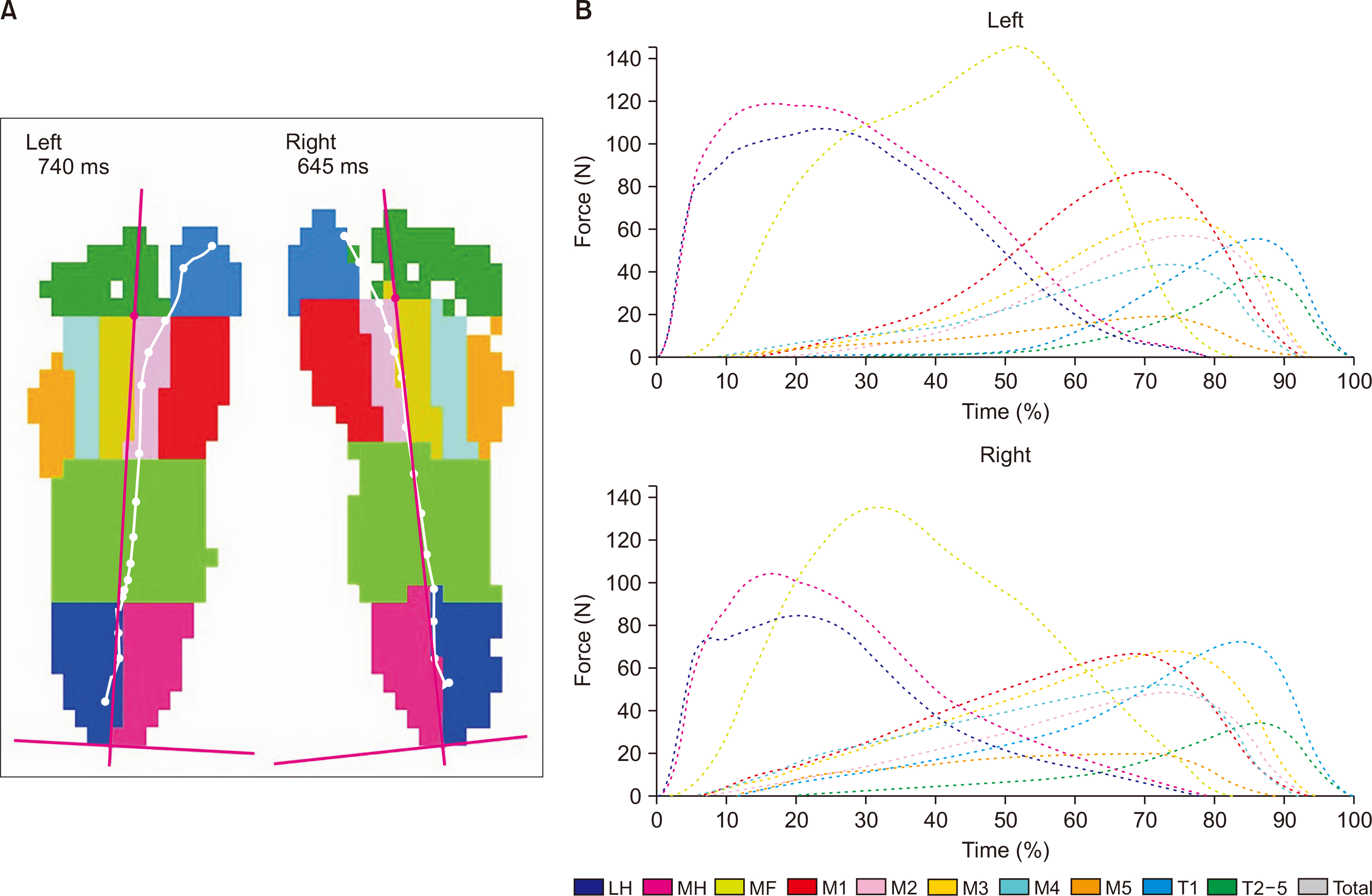
Figure. 2 Pedobarographic result. (A) Ten anatomical zones. (B) Graph shows the ten anatomical zones analysis of the dynamic recording.
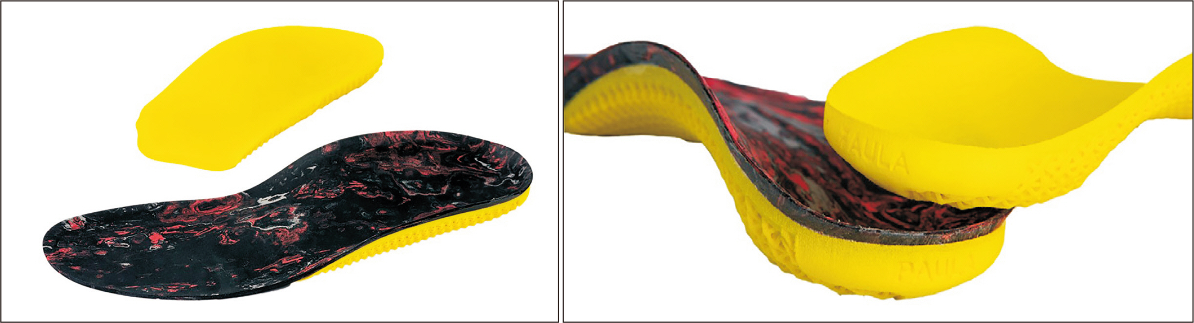
Figure. 3 Three-dimensional printing insole.
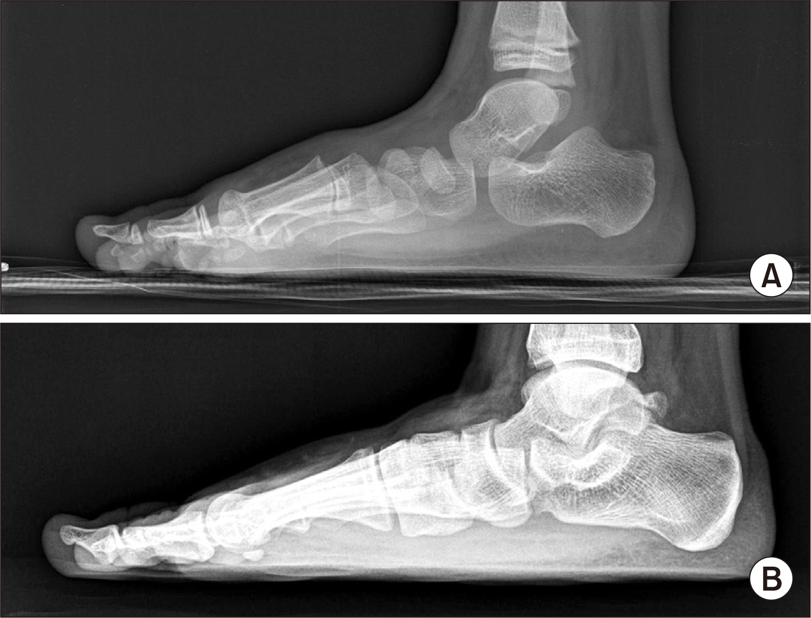
Figure. 4 Initial (A) and last follow-up (B) radiograph. Radiographic parameters were improved in last follow-up radiograph. But all values were still within the abnormal range.
Cited by 1 articles
-
Change in Plantar Pressure and Plain Radiography in Pediatric Flexible Flatfoot: A Retrospective Cohort Study
Sungjoon Kim, Yong Gyun Kim, Jun Yup Kim, Si-Bog Park, Kyu Hoon Lee
Ann Rehabil Med. 2024;48(5):352-359. doi: 10.5535/arm.240041.
Reference
-
1. Staheli LT, Chew DE, Corbett M. 1987; The longitudinal arch. A survey of eight hundred and eighty-two feet in normal children and adults. J Bone Joint Surg Am. 69:426–8. DOI: 10.2106/00004623-198769030-00014. PMID: 3818704.2. Vanderwilde R, Staheli LT, Chew DE, Malagon V. 1988; Measurements on radiographs of the foot in normal infants and children. J Bone Joint Surg Am. 70:407–15. DOI: 10.2106/00004623-198870030-00013. PMID: 3346265.
Article3. Bok SK, Kim BO, Lim JH, Ahn SY. 2014; Effects of custom-made rigid foot orthosis on pes planus in children over 6 years old. Ann Rehabil Med. 38:369–75. doi: 10.5535/arm.2014.38.3.369. DOI: 10.5535/arm.2014.38.3.369. PMID: 25024961. PMCID: PMC4092178.
Article4. Bordelon RL. 1980; Correction of hypermobile flatfoot in children by molded insert. Foot Ankle. 1:143–50. doi: 10.1177/107110078000100303. DOI: 10.1177/107110078000100303. PMID: 7319430.
Article5. Choi JY, Hong WH, Suh JS, Han JH, Lee DJ, Lee YJ. 2020; The long-term structural effect of orthoses for pediatric flexible flat foot: a systematic review. Foot Ankle Surg. 26:181–8. doi: 10.1016/j.fas.2019.01.007. DOI: 10.1016/j.fas.2019.01.007. PMID: 30765257.
Article6. Gould N, Moreland M, Alvarez R, Trevino S, Fenwick J. 1989; Development of the child's arch. Foot Ankle. 9:241–5. doi: 10.1177/107110078900900506. DOI: 10.1177/107110078900900506. PMID: 2731836.
Article7. Kanatlı U, Aktas E, Yetkin H. 2016; Do corrective shoes improve the development of the medial longitudinal arch in children with flexible flat feet? J Orthop Sci. 21:662–6. doi: 10.1016/j.jos.2016.04.014. DOI: 10.1016/j.jos.2016.04.014. PMID: 27212230.
Article8. Mereday C, Dolan CM, Lusskin R. 1972; Evaluation of the University of California Biomechanics Laboratory shoe insert in “flexible” pes planus. Clin Orthop Relat Res. 82:45–58. DOI: 10.1097/00003086-197201000-00006. PMID: 5011037.
Article9. Riccio I, Gimigliano F, Gimigliano R, Porpora G, Iolascon G. 2009; Rehabilitative treatment in flexible flatfoot: a perspective cohort study. Chir Organi Mov. 93:101–7. doi: 10.1007/s12306-009-0037-z. DOI: 10.1007/s12306-009-0037-z. PMID: 19777377.
Article10. Wenger DR, Mauldin D, Speck G, Morgan D, Lieber RL. 1989; Corrective shoes and inserts as treatment for flexible flatfoot in infants and children. J Bone Joint Surg Am. 71:800–10. DOI: 10.2106/00004623-198971060-00002. PMID: 2663868.
Article11. Seol YJ, Jung ST, Yang HK, Lee KB, Oh CS, Jung YJ, et al. 2014; Diagnostic availability of pedobarography and correlation of radiographic and pedobarographic measurements in pediatric flexible flatfoot. J Korean Orthop Assoc. 49:366–73. doi: 10.4055/jkoa.2014.49.5.366. DOI: 10.4055/jkoa.2014.49.5.366.
Article12. Erdemir A, Saucerman JJ, Lemmon D, Loppnow B, Turso B, Ulbrecht JS, et al. 2005; Local plantar pressure relief in therapeutic footwear: design guidelines from finite element models. J Biomech. 38:1798–806. doi: 10.1016/j.jbiomech.2004.09.009. DOI: 10.1016/j.jbiomech.2004.09.009. PMID: 16023466.
Article13. Telfer S, Gibson KS, Hennessy K, Steultjens MP, Woodburn J. 2012; Computer-aided design of customized foot orthoses: reproducibility and effect of method used to obtain foot shape. Arch Phys Med Rehabil. 93:863–70. doi: 10.1016/j.apmr.2011.12.019. DOI: 10.1016/j.apmr.2011.12.019. PMID: 22541310.
Article14. Cha YH, Lee KH, Ryu HJ, Joo IW, Seo A, Kim DH, et al. 2017; Ankle-foot orthosis made by 3D printing technique and automated design software. Appl Bionics Biomech. 2017:9610468. doi: 10.1155/2017/9610468. DOI: 10.1155/2017/9610468. PMID: 28827977. PMCID: PMC5554564.
Article15. Xu R, Wang Z, Ma T, Ren Z, Jin H. 2019; Effect of 3D printing individualized ankle-foot orthosis on plantar biomechanics and pain in patients with plantar fasciitis: a randomized controlled trial. Med Sci Monit. 25:1392–400. doi: 10.12659/MSM.915045. DOI: 10.12659/MSM.915045. PMID: 30789873. PMCID: PMC6394143.
Article16. Mickle KJ, Steele JR, Munro BJ. 2008; Is the foot structure of preschool children moderated by gender? J Pediatr Orthop. 28:593–6. doi: 10.1097/BPO.0b013e318173f782. DOI: 10.1097/BPO.0b013e318173f782. PMID: 18580378.
Article17. Banwell HA, Paris ME, Mackintosh S, Williams CM. 2018; Paediatric flexible flat foot: how are we measuring it and are we getting it right? A systematic review. J Foot Ankle Res. 11:21. doi: 10.1186/s13047-018-0264-3. DOI: 10.1186/s13047-018-0264-3. PMID: 29854006. PMCID: PMC5975578.
Article18. Bok SK, Lee H, Kim BO, Ahn S, Song Y, Park I. 2016; The effect of different foot orthosis inverted angles on plantar pressure in children with flexible flatfeet. PLoS One. 11:e0159831. doi: 10.1371/journal.pone.0159831. DOI: 10.1371/journal.pone.0159831. PMID: 27458719. PMCID: PMC4961415.
Article19. Rose GK, Welton EA, Marshall T. 1985; The diagnosis of flat foot in the child. J Bone Joint Surg Br. 67:71–8. doi: 10.1302/0301-620X.67B1.3968149. DOI: 10.1302/0301-620X.67B1.3968149. PMID: 3968149.
Article20. Joo SY, Kim JR. 2016; Management of flexible flatfoot in chidren and adolescent. J Korean Orthop Assoc. 51:109–16. doi: 10.4055/jkoa.2016.51.2.109. DOI: 10.4055/jkoa.2016.51.2.109.
Article21. Carr JB 2nd, Yang S, Lather LA. 2016; Pediatric pes planus: a state-of-the-art review. Pediatrics. 137:e20151230. doi: 10.1542/peds.2015-1230. DOI: 10.1542/peds.2015-1230. PMID: 26908688.
Article22. Kuhn DR, Shibley NJ, Austin WM, Yochum TR. 1999; Radiographic evaluation of weight-bearing orthotics and their effect on flexible pes planus. J Manipulative Physiol Ther. 22:221–6. doi: 10.1016/s0161-4754(99)70048-5. DOI: 10.1016/S0161-4754(99)70048-5. PMID: 10367758.
Article23. Kitaoka HB, Luo ZP, Kura H, An KN. 2002; Effect of foot orthoses on 3-dimensional kinematics of flatfoot: a cadaveric study. Arch Phys Med Rehabil. 83:876–9. doi: 10.1053/apmr.2002.32681. DOI: 10.1053/apmr.2002.32681. PMID: 12048672.
Article24. Ferri M, Scharfenberger AV, Goplen G, Daniels TR, Pearce D. 2008; Weightbearing CT scan of severe flexible pes planus deformities. Foot Ankle Int. 29:199–204. doi: 10.3113/FAI.2008.0199. DOI: 10.3113/FAI.2008.0199. PMID: 18315976.
Article25. Choi JY, Lee DJ, Kim SJ, Suh JS. 2020; Does the long-term use of medial arch support insole induce the radiographic structural changes for pediatric flexible flat foot?- A prospective comparative study. Foot Ankle Surg. 26:449–56. doi: 10.1016/j.fas.2019.05.017. DOI: 10.1016/j.fas.2019.05.017. PMID: 31201010.26. Aboutorabi A, Saeedi H, Kamali M, Farahmand B, Eshraghi A, Dolagh RS. 2014; Immediate effect of orthopedic shoe and functional foot orthosis on center of pressure displacement and gait parameters in juvenile flexible flat foot. Prosthet Orthot Int. 38:218–23. doi: 10.1177/0309364613496111. DOI: 10.1177/0309364613496111. PMID: 23986466.
Article
- Full Text Links
-
- Actions
-
Cited
- CITED
-
- Close
- Share
-
- Similar articles
-
- The Effect of Insole to Flexible Flat Foot on Dynamic Balance and Ankle Muscle Activity during the Y-Balance Test
- Comparison of the Forefoot Pressure-Relieving Effects of Foot Orthoses
- Effects of three-dimensional image based insole for healthy volunteers: a pilot clinical trial
- The Effect of Metatarsal Pad for Foot Pressure
- The Treatment of Failed Kidner Procedure for Adolescent Prehallux: A Case Report
