Neurointervention.
2024 Nov;19(3):185-189. 10.5469/neuroint.2024.00318.
Delayed Pontomesencephalic and Cervical Cord Venous Drainage Followed by Contralateral Carotid-Cavernous Fistula after Craniofacial Fractures: A Case Report
- Affiliations
-
- 1Department of Neurosurgery, Faculty of Medicine, Universitas Sumatera Utara, Medan, Indonesia
- 2Department of Neurosurgery, Siloam Dhirga Surya Hospital, Medan, Indonesia
- 3Faculty of Medicine, Pelita Harapan University, Tangerang, Indonesia
- 4Department of Radiology, Siloam Dhirga Surya Hospital, Medan, Indonesia
- KMID: 2560224
- DOI: http://doi.org/10.5469/neuroint.2024.00318
Abstract
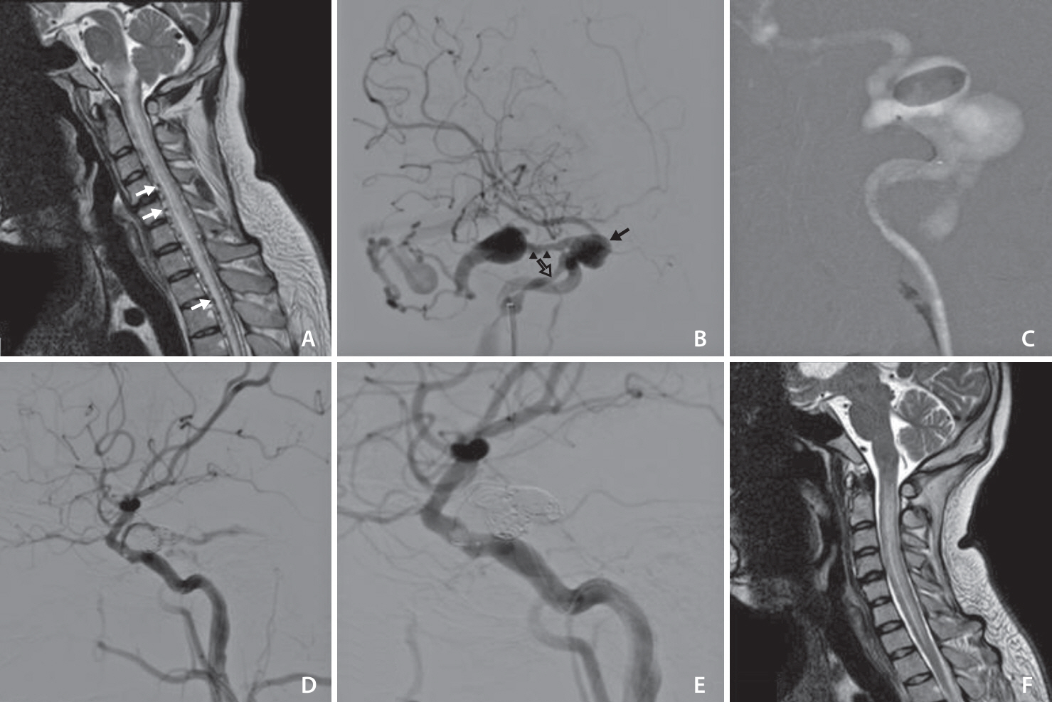
- A 24-year-old male was admitted with progressive cervical hypesthesia, tetraparesis, dyspnea, and a history of craniofacial fracture. Spinal magnetic resonance imaging (MRI) showed brainstem edema extending to the thoracic spine with multiple prominent perimedullary vascular structures. Cerebral digital-substraction angiography revealed Barrow type A carotid-cavernous fistula. Total occlusion with preservation of internal carotid artery flow was achieved using 1 detachable balloon and 6 coils. Postoperatively, immediate respiratory recovery, gradual extremities strength improvement, and right abducens nerve palsy were found. One month follow-up cervical MRI showed good recovery of spinal cord edema and perimedullary veins.
Figure
Reference
-
1. Barrow DL, Spector RH, Braun IF, Landman JA, Tindall SC, Tindall GT. Classification and treatment of spontaneous carotid-cavernous sinus fistulas. J Neurosurg. 1985; 62:248–256.2. Xu XQ, Liu S, Zu QQ, Zhao LB, Xia JG, Zhou CG, et al. Follow-up of 58 traumatic carotid-cavernous fistulas after endovascular detachable-balloon embolization at a single center. J Clin Neurol. 2013; 9:83–90.3. Henderson AD, Miller NR. Carotid-cavernous fistula: current concepts in aetiology, investigation, and management. Eye (Lond). 2018; 32:164–172.4. Korkmazer B, Kocak B, Tureci E, Islak C, Kocer N, Kizilkilic O. Endovascular treatment of carotid cavernous sinus fistula: a systematic review. World J Radiol. 2013; 5:143–155.5. Narita Y, Watanabe Y, Hoshino T, Okada M, Yamamoto Y, Kuzuhara S. Myelopathy due to large veins draining recurrent spontaneous caroticocavernous fistula. Neuroradiology. 1992; 34:433–435.6. Ko SB, Kim CK, Lee SH, Yoon BW. Carotid cavernous fistula with cervical myelopathy. J Clin Neurosci. 2009; 16:1350–1353.7. Herrera DA, Vargas SA, Dublin AB. Traumatic carotid-cavernous fistula with pontomesencephalic and cervical cord venous drainage presenting as tetraparesis. J Neuroimaging. 2011; 21:73–75.8. Ding CL, Zhang CL, Hua F, Xi SD, Zhou QW, Wang HJ, et al. Traumatic carotid-cavernous fistula with perimedullary venous drainage and delayed myelopathy: A case report. Med Int (Lond). 2021; 1:16.9. Ract I, Drier A, Leclercq D, Sourour N, Gabrieli J, Yger M, et al. Extensive basal ganglia edema caused by a traumatic carotid-cavernous fistula: a rare presentation related to a basal vein of Rosenthal anatomical variation. J Neurosurg. 2014; 121:63–66.10. Ma YH, Shang R, Lin S, Li SH, Wang T, Zhang CW. Case report: delayed quadriplegia from traumatic carotid cavernous fistula: a rare case with perimedullary venous drainage. Front Neurol. 2023; 14:1224425.11. Ellis JA, Goldstein H, Connolly ES Jr, Meyers PM. Carotid-cavernous fistulas. Neurosurg Focus. 2012; 32:E9.12. Ying Sze C, Bakin S, Ismail R. Neuropsychiatric presentation in indirect carotid-cavernous fistula: a case report. Cureus. 2023; 15:e37523.13. Aralasmak A, Karaali K, Cevikol C, Senol U, Sindel T, Toprak H, et al. Venous drainage patterns in carotid cavernous fistulas. ISRN Radiol. 2014; 2014:760267.14. Naylor RM, Topinka B, Rinaldo L, Jacobi J, Neth B, Flemming KD, et al. Progressive myelopathy from a craniocervical junction dural arteriovenous fistula. Stroke. 2021; 52:e278–e281.15. Kim WY, Kim JB, Nam TK, Kim YB, Park SW. Cervical myelopathy caused by intracranial dural arteriovenous fistula. Korean J Spine. 2016; 13:67–70.16. D’Angelo L, Paglia F, Caporlingua A, Sampirisi L, Guidetti G, Santoro A. Atypical manifestation of direct low-flow carotid-cavernous fistula: case report and review of the literature. World Neurosurg. 2019; 125:456–460.17. Shim HS, Kang KJ, Choi HJ, Jeong YJ, Byeon JH. Delayed contralateral traumatic carotid cavernous fistula after craniomaxillofacial fractures. Arch Craniofac Surg. 2019; 20:44–47.18. Ide S, Kiyosue H, Tokuyama K, Hori Y, Sagara Y, Kubo T. Direct carotid cavernous fistulas. J Neuroendovasc Ther. 2020; 14:583–592.19. Cunningham EJ, Albani B, Masaryk TJ, Rasmussen PA. Temporary balloon occlusion of the cavernous carotid artery for removal of an orbital and intracranial foreign body: case report. Neurosurgery. 2004; 55:1225.20. Fukawa N, Nakagawa N, Tsuji K, Yoshioka H, Furukawa K, Nagatsuka K, et al. Transarterial and transvenous coil embolization of direct carotid-cavernous fistulas. J Neuroendovasc Ther. 2022; 16:127–134.21. Yu J, Guo Y, Zhao S, Xu K. Brainstem edema caused by traumatic carotid-cavernous fistula: a case report and review of the literature. Exp Ther Med. 2015; 10:445–450.
- Full Text Links
- Actions
-
Cited
- CITED
-
- Close
- Share
- Similar articles
-
- Traumatic Carotid-cavernous Fistula with Contralateral Proptosis: Case Report
- Delayed contralateral traumatic carotid cavernous fistula after craniomaxillofacial fractures
- Traumatic Carotid-cavernous Fistula Bringing about Intracerebral Hemorrhage
- Delayed intracerebral hemorrhage from a traumatic carotid-cavernous fistula associated with an enucleated orbit
- A Case of Dural Carotid-Cavernous Sinus Fistula Associated with Ophthalmic Manifestations


